Patterson’s Curse Pyrrolizidine-Alkaloid Poisoning, Cumulative Liver Failure, Photosensitization, and Hepatic Encephalopathy
Is Patterson’s Curse Poisonous to Dogs, Cats, Horses, and Livestock?
Yes—Patterson’s Curse, Echium plantagineum L., is poisonous to horses, pigs, cattle, sheep, goats, donkeys, camelids, and other grazing animals, and it should also be kept away from dogs, cats, rabbits, guinea pigs, companion birds, poultry, reptiles, and other animals. The standard Australian spelling is Paterson’s Curse, with one “t,” while Patterson’s Curse is a recognized search and title variant. Rosette leaves, flowering stems, flowers, roots, developing fruit, rough nutlets and their seeds, dead plants, and material incorporated into hay, silage, pellets, grain, bedding, crop residue, or herbal products should all be treated as potentially toxic.
The plant contains hepatotoxic 1,2-unsaturated pyrrolizidine alkaloids and their N-oxides. After absorption, liver enzymes can convert these compounds into highly reactive dehydropyrrolizidine metabolites that bind to DNA, proteins, and other cellular structures. Repeated injury interferes with normal liver-cell division and regeneration, producing megalocytosis, individual-cell necrosis, bile-duct proliferation, fibrosis, cirrhosis, portal hypertension, impaired detoxification, and eventual liver failure.
Poisoning is commonly delayed and cumulative. An animal may continue eating and appear outwardly healthy while microscopic damage develops over weeks, months, or more than one grazing season, and disease can continue progressing after the contaminated pasture or feed has been removed. Early signs may be limited to reduced appetite, poor performance, dullness, sleepiness, weight loss, reduced wool or growth production, or separation from the herd before jaundice, ascites, photosensitization, difficulty swallowing, hepatic encephalopathy, collapse, or death becomes apparent.
Horses and pigs are among the most susceptible domestic animals, while cattle remain vulnerable and sheep and goats are comparatively resistant rather than immune. Sheep with PA-damaged livers may retain excessive copper and later suffer a sudden hemolytic crisis marked by weakness, jaundice, rapid breathing, anemia, and dark red-brown urine. Dogs, cats, rabbits, and birds are less commonly documented because ordinary household exposure is usually brief, but repeated plant ingestion, contaminated forage, seed-contaminated feed, or a large acute dose still requires veterinary evaluation.
About this guide: This page provides general pet-poisoning information and cannot diagnose or treat an individual animal. For any suspected exposure, contact a veterinarian or animal poison-control service immediately. Do not induce vomiting, give medication, or attempt home decontamination unless directed by a veterinary professional.
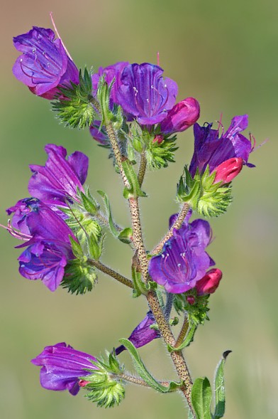
Patterson's Curse
Echium plantagineum L.
- Echium creticum subsp. plantagineum (L.) Malag. — homotypic historical subspecies combination
- Echium lycopsis L. — rejected historical name widely encountered in older botanical literature
- Echium violaceum L. — historical heterotypic synonym
- Echium bonariense Poir. — historical heterotypic synonym
- Echium longistamineum Pourr. ex Lapeyr. — historical heterotypic synonym
- Echium maritimum Willd. — historical heterotypic synonym
- Echium orientale Stephan — historical heterotypic synonym
- Echium plantaginifolium L. ex Moris — historical spelling and nomenclatural variation
- Echium plantaginoides Roem. & Schult. — historical heterotypic synonym
- Echium pseudoviolaceum Schur — historical heterotypic synonym
Boraginaceae — Borage or Forget-Me-Not Family
Patterson’s Curse; Paterson’s Curse; Paterson Curse; Salvation Jane; Purple Viper’s-Bugloss; Purple Viper's Bugloss; Plantain-Leaved Viper’s-Bugloss; Plantain Viper’s-Bugloss; Riverina Bluebell; Blueweed; Lady Campbell Weed; Purple Echium
Historical and taxonomic search variations include Echium creticum subsp. plantagineum (L.) Malag., Echium lycopsis L., Echium violaceum L., Echium bonariense Poir., Echium longistamineum Pourr. ex Lapeyr., Echium maritimum Willd., Echium orientale Stephan, Echium plantaginifolium L. ex Moris, Echium plantaginoides Roem. & Schult., and Echium pseudoviolaceum Schur.
Viper’s Bugloss without the modifiers “purple” or “plantain-leaved” more commonly refers to the separate species Echium vulgare, which is also a bristly, blue-purple-flowered member of Boraginaceae and may contain hepatotoxic pyrrolizidine alkaloids. Echium plantagineum usually has broad plantain-like rosette leaves with conspicuous branched lateral veins, a comparatively long purple funnel-shaped corolla, and two stamens projecting beyond the flower, while E. vulgare generally has narrower leaves, a shorter corolla, and four conspicuously exserted stamens. The scientific name, complete rosette, stem leaves, flower tube, protruding stamens, rough four-part nutlet cluster, geographic location, and forage source should be used for identification.
Hepatotoxic 1,2-Unsaturated Pyrrolizidine Alkaloids
Patterson’s Curse contains a chemically complex mixture of pyrrolizidine alkaloids and pyrrolizidine-alkaloid N-oxides. The toxicologically important structures possess a 1,2-unsaturated necine base that can undergo hepatic metabolic activation. Saturation, ester structure, stereochemistry, tissue concentration, and the attached necic acids influence activity, so the term pyrrolizidine alkaloid describes a large chemical class rather than one uniform poison.
Modern exact-species analytical work has detected numerous alkaloids and N-oxides in Echium plantagineum. Reported compounds include echimidine, echiumine, echihumiline, hydroxymyoscorpine, lycopsamine, intermedine, related acetylated structures, leptanthine-related material, and multiple isomers whose chromatographic behavior can make exact identification difficult. Not every reported compound occurs at the same concentration in every population, tissue, growth stage, or analytical sample.
The plant often contains a substantial proportion of its alkaloid burden as N-oxides. N-oxide status does not make the plant harmless because digestive and metabolic processes can reduce these compounds to tertiary alkaloids capable of subsequent activation. Feed analysis that measures only free-base alkaloids may therefore underestimate the relevant total PA burden.
Echimidine, Echihumiline, and Hydroxymyoscorpine
Echimidine has long been treated as a principal or characteristic alkaloid of Patterson’s Curse. Recent separation and nuclear-magnetic-resonance work demonstrated that an apparent echimidine peak produced under common acidic HPLC conditions can contain echimidine together with its C-7 isomers echihumiline and hydroxymyoscorpine. Earlier analytical reports may therefore have combined several structures under one apparent echimidine measurement.
Exact samples collected in New Zealand, Argentina, and the United States showed similar co-occurrence of the three isomeric compounds. Before chemical reduction, much of the echimidine and echihumiline material in the examined sample occurred as N-oxides. These findings improve chemical accuracy but do not provide a validated plant percentage or blood concentration that predicts disease in an individual horse, cow, pig, sheep, dog, or bird.
In primary rat-hepatocyte cultures, echimidine and the combined isomer fraction produced concentration-dependent loss of cell viability. The experimental concentrations and direct cell exposure do not reproduce grazing, ruminal transformation, gastrointestinal absorption, whole-animal metabolism, or chronic natural feeding. The study confirms intrinsic hepatotoxic potential while leaving veterinary toxicokinetics and species-specific dose response unresolved.
Echiumine Is a Separate Alkaloid
Echiumine and echimidine are separate pyrrolizidine alkaloids and should not be treated as alternate spellings. Both were isolated historically from Echium plantagineum, and each possesses a retronecine-based structure with different esterifying components. The distinction matters because analytical reports and secondary summaries sometimes merge the names.
Echiumine produced greater toxicity than echimidine or the echimidine-isomer fraction in the modern rat-hepatocyte comparison. That result concerns one experimental model and should not be converted directly into the claim that echiumine is always the principal cause of livestock death. Natural tissues expose animals to mixtures whose concentrations, absorption, and metabolic activation differ.
Absorption and Hepatic Metabolic Activation
Ingested alkaloids and reducible N-oxides are absorbed through the gastrointestinal tract and carried through portal blood toward the liver. Hepatic cytochrome-P450 enzymes can participate in detoxification pathways, but they can also oxidize toxic unsaturated PAs into unstable dehydropyrrolizidine intermediates commonly described as reactive pyrrolic metabolites. The balance between detoxification and activation varies by compound, dose, species, age, enzyme activity, gut microorganisms, and nutritional state.
Reactive metabolites can bind covalently to cellular proteins, DNA, nucleoproteins, enzymes, and membrane-associated structures. Some metabolites are hydrolyzed or conjugated and eliminated, while others form persistent adducts that interfere with cell function and repair. The animal is therefore injured not because unchanged plant alkaloid simply accumulates forever in the liver, but because metabolism creates molecular damage that may remain after the original exposure has ended.
Extrahepatic injury is possible when reactive metabolites or more stable secondary products reach other tissues, but the liver remains the principal target because it receives portal blood and performs the activating metabolism. Lung, vascular, kidney, and other lesions occur with some PAs and species, yet chronic hepatic failure is the defining veterinary concern for Patterson’s Curse. Atypical organ disease still requires investigation for another plant, compound, or concurrent illness.
DNA and Protein Adducts
Reactive pyrrolic metabolites can cross-link DNA, DNA-associated proteins, and other cellular macromolecules. This disrupts replication, transcription, repair, and ordinary cell-cycle progression. Pyrrole-protein and pyrrole-DNA adducts may persist longer than the freely circulating parent alkaloids and can provide evidence of earlier exposure when specialized testing is available.
Adduct detection is not a routine field-side test and must be interpreted with the specimen type, laboratory method, elapsed time, and competing PA sources. A positive result supports exposure to metabolically activated pyrrolizidine alkaloids but may not identify the exact plant without botanical and feed evidence. A negative result does not always exclude remote or low-level exposure.
Megalocytosis and Failure of Liver Regeneration
One characteristic result of pyrrolizidine-alkaloid injury is hepatocellular megalocytosis. Damaged hepatocytes continue synthesizing cellular material and enlarging, but they cannot complete normal cell division. Their nuclei and cytoplasm become markedly enlarged while the liver loses its ordinary regenerative response.
A healthy liver can compensate for substantial injury by replacing lost cells. PA-damaged liver tissue cannot regenerate efficiently because the surviving cells themselves have been rendered antimitotic. Continued individual-cell death therefore removes functional tissue faster than it can be replaced.
Megalocytosis is an important histopathologic clue but is not completely exclusive to one plant or one PA. It must be interpreted with fibrosis, bile-duct changes, exposure history, and the distribution of lesions. Small or early biopsies may underestimate disease because damage can vary within the organ.
Necrosis, Bile-Duct Proliferation, and Progressive Fibrosis
Repeated toxic injury causes individual hepatocyte necrosis, inflammatory responses, bile-duct proliferation, nodular regeneration, distortion of lobular architecture, and increasing deposition of fibrous connective tissue. Surviving tissue becomes divided and compressed by scar. Blood and bile can no longer move through the organ normally.
Fibrosis increases resistance to portal blood flow and contributes to portal hypertension. Albumin production falls, bilirubin clearance becomes inadequate, clotting-factor production can decline, and gastrointestinally derived toxins are no longer removed effectively. These changes explain the eventual combination of weight loss, jaundice, ascites, edema, bleeding tendency, and hepatic encephalopathy.
Advanced fibrosis and cirrhosis are not reversible through charcoal, vitamins, herbs, amino-acid supplements, or short-term dietary manipulation. Removing exposure protects remaining tissue but does not recreate hepatocytes that have been lost or trapped within severe scar. Treatment must work with whatever functional liver reserve remains.
Cumulative Injury and Delayed Disease
Repeated individually small doses can produce severe cumulative disease. The animal may graze the weed intermittently, consume scattered fragments in hay, or receive contaminated feed for weeks without immediate illness. Damage continues silently until functional reserve falls below the level needed to maintain normal metabolism.
Clinical signs may appear after the weed has died, the pasture has changed, or the contaminated hay lot has been exhausted. This delay can obscure the true source and falsely implicate a new ration or recent event. Exposure histories must cover previous grazing seasons, former paddocks, purchased forage, crop residues, and earlier feed batches.
Disease may continue progressing after complete removal because previously injured hepatocytes continue dying and scar formation continues. Removal is nevertheless essential because every additional dose reduces the chance that enough functional tissue will survive. Follow-up testing must continue after the source has been eliminated.
Acute High-Dose Hepatotoxicity
Chronic cumulative disease is the usual presentation, but a sufficiently large exposure can produce acute or subacute hepatic necrosis. Experimental feeding of young rats produced fatal hemorrhagic liver injury after comparatively short exposure periods, and continuing deaths occurred after the toxic diet had stopped. Acute cases may develop severe depression, abdominal pain, weakness, hemorrhage, rapid loss of liver function, collapse, and death before classic chronic fibrosis is fully expressed.
Acute presentation does not make home gastrointestinal decontamination appropriate after clinical liver failure has developed. By that stage the relevant compounds have generally been absorbed and metabolically activated. Emergency care must address circulation, glucose, coagulation, neurologic function, and organ failure while the source is removed from every exposed animal.
Genotoxicity and Carcinogenic Potential
Reactive PA metabolites can damage DNA and have produced mutations, chromosomal injury, and tumors in experimental systems. The carcinogenic potential is relevant to food-chain control and long-term public-health regulation. It is not usually the first clinical problem recognized in severely poisoned livestock because progressive liver failure may develop earlier.
A surviving animal with subclinical exposure cannot be assumed free of long-term molecular damage. Food-producing-animal decisions may require veterinary, regulatory, residue, welfare, and market considerations beyond the treatment of visible signs. Milk, meat, eggs, honey, and other animal products should not be assessed casually after a significant exposure.
Root Naphthoquinones and the Toxicology Boundary
Exact metabolic profiling has identified shikonin and related naphthoquinones in Patterson’s Curse roots in addition to pyrrolizidine-alkaloid N-oxides. These compounds contribute to plant defense, antimicrobial effects, allelopathy, and the ecological success of the weed. Their presence expands the plant’s chemical profile beyond PAs.
Shikonins have biological activity in experimental systems, but they have not been established as the principal cause of the characteristic chronic livestock liver syndrome. Public veterinary guidance should therefore retain 1,2-unsaturated pyrrolizidine alkaloids as the primary toxic mechanism. Roots remain unsuitable for animal access because they contain biologically active plant chemistry and may be contaminated with soil treatments or herbicides.
All Plant Parts and Processed Feed
Rosette leaves, stem leaves, flowering shoots, flowers, roots, developing fruit, rough nutlets, seeds, senescent plants, and dead material should all be treated as potentially toxic. Alkaloid concentration may differ among those tissues, but no tissue has been established as universally safe. Pulled or cut flowering plants may continue maturing nutlets and adding viable seed to the environment.
Drying does not reliably destroy pyrrolizidine alkaloids. Hay can be especially hazardous because bristly texture and poor palatability no longer allow animals to avoid the plant selectively, while chopped or baled fragments are difficult to recognize. Pelleting, grinding, ensiling, mixing into grain, or incorporation into crop residue can distribute small repeated doses throughout the ration.
Bedding contamination also matters when horses, pigs, rabbits, or other animals eat bedding material. Seed-contaminated grain and rejected cleaning fractions may introduce material into poultry or pig feed. One clean-looking handful does not clear an entire bale, silo, bin, truckload, or batch.
Seasonal, Geographic, and Environmental Variation
Alkaloid profiles differ among geographic populations. Exact sampling across southeastern Australia found consistent PA and N-oxide occurrence but meaningful differences in abundance among locations. Climate, latitude, soil, plant genetics, drought, temperature, growth stage, herbivory, and other stresses may alter secondary-metabolite production.
Reports disagree about whether rosette or flowering plants contain the highest total alkaloid burden. The disagreement likely reflects different populations, tissues, seasons, and analytical methods rather than proof that one official source is universally wrong. Animals must not be used to identify a safe stage through trial grazing.
Flowering and seed production still increase practical exposure because the plant becomes conspicuous, can dominate pasture, contaminates harvested forage, and adds many rough nutlets to the soil and feed chain. Rosette-stage plants may be more palatable and can contain substantial alkaloid concentrations. Every stage requires control.
Species Susceptibility
Horses and pigs are considered highly susceptible. As nonruminants, they lack the prolonged foregut fermentation that can transform part of the PA burden before absorption, although individual metabolic differences remain important. Horses also commonly develop dramatic hepatic encephalopathy once liver reserve is exhausted.
Cattle possess ruminal microorganisms capable of reducing part of the effective dose but remain moderately susceptible under sustained exposure. Chronic outbreaks have produced weight loss, diarrhea, jaundice, photosensitization, fibrosis, and death. Rumen function reduces risk; it does not neutralize a heavily contaminated ration.
Sheep and goats are comparatively resistant because of ruminal metabolism, species-specific detoxification, and grazing behavior. Resistance varies by breed, individual, previous exposure, rumen population, diet, copper intake, and management. Sheep may develop PA-associated liver damage and dangerous copper retention even when ordinary serum enzymes or outward appearance remain unremarkable.
Dogs, cats, rabbits, birds, reptiles, and other companion animals have not been studied as extensively with this exact plant. Their risk depends on the dose and duration they actually receive, not on the absence of published case series. Repeated access to contaminated forage, dried herbs, seed material, or a dense infestation should be treated seriously.
Young and Growing Animals
Young animals may be particularly vulnerable because their hepatocytes are actively dividing and because small body size can produce a larger dose per unit of body weight. Experimental young rats developed extensive injury and high mortality after shorter exposure than would be expected from the plant’s reputation as only a slow chronic poison. The exact age effect in each domestic species remains incompletely quantified.
Growing livestock may first show reduced weight gain, delayed development, or lower production rather than jaundice. Those changes can be mistaken for parasites, poor forage quality, mineral imbalance, or infectious disease. Group records and feed history are therefore important even before obvious liver failure appears.
No Dependable Safe Dose
No universal safe plant percentage, daily intake, nutlet count, grazing duration, bale contamination level, or body-weight dose applies to every animal. Alkaloid concentration and metabolic susceptibility vary too widely. One horse may become ill after several weeks of dense grazing while another exposed in the same paddock may not show disease until a later season.
A low measured concentration in one sample does not guarantee that every portion of the pasture or feed lot is equally low. Plant fragments may be distributed unevenly, and a few bales can contain much greater contamination than the average. Risk assessment requires representative sampling and veterinary interpretation rather than visual guesswork.
Delayed and Initially Invisible Disease
Patterson’s Curse poisoning is usually chronic, cumulative, and clinically silent during its early development. Animals may graze an infested paddock or consume contaminated hay for weeks or months while microscopic hepatocyte injury, megalocytosis, bile-duct proliferation, and fibrosis accumulate. Visible illness often begins only after a substantial part of the liver’s functional reserve has been lost.
The weed may no longer be present when disease becomes obvious. Pasture rotation, drought, seasonal dieback, hay-lot changes, sale between properties, or removal of an obvious infestation can separate the exposure from the eventual diagnosis. Historical feed and grazing records may therefore be more valuable than inspection of the current paddock alone.
Removal from the source does not guarantee immediate stabilization. Previously damaged cells can continue dying and scar can continue distorting the remaining organ. Animals without visible signs may still require repeated testing over an extended period.
Early Nonspecific Changes
Early clinical changes commonly include dullness, sleepiness, reduced appetite, slower eating, diminished performance, poor growth, progressive weight loss, and declining body condition. An affected animal may separate from the group, spend more time standing quietly, or appear less responsive to ordinary activity. Horses may become exercise intolerant, while production animals may grow less wool, gain less weight, or show reduced milk or reproductive performance.
These findings overlap with parasites, dental disease, inadequate nutrition, chronic infection, gastrointestinal disease, and many other disorders. An apparently reasonable ration does not exclude toxic contamination. The presence of Patterson’s Curse in pasture or harvested feed must be investigated directly.
Jaundice and Bilirubin Accumulation
Jaundice may become visible in the gums, conjunctiva, sclera, vulva, unpigmented skin, or other lightly colored tissues. It develops when the damaged liver cannot process and excrete bilirubin normally or when hemolysis adds a large bilirubin load. Color can range from faint yellowing to intense orange-yellow discoloration.
Jaundice indicates significant hepatic dysfunction or red-blood-cell destruction and should never be treated as a minor pasture reaction. It may be accompanied by dark urine, pale feces, weakness, photosensitization, bleeding tendency, or neurologic disease. Laboratory testing is required to distinguish primary hepatic failure from a secondary copper-associated hemolytic crisis.
Ascites, Edema, and Abdominal Enlargement
The abdomen may enlarge because yellowish fluid accumulates within the peritoneal cavity. Portal hypertension impedes normal venous flow through the fibrotic liver, while reduced albumin production lowers the plasma’s ability to retain fluid. Both mechanisms can contribute simultaneously.
Dependent edema may develop beneath the chest, abdomen, jaw, or limbs. A distended abdomen can impair comfort, movement, breathing, and appetite. Enlargement must be distinguished from pregnancy, obesity, parasitism, gastrointestinal distension, bladder disease, hemorrhage, and other causes.
Ascites does not mean that uncontrolled drainage is automatically appropriate. Removing fluid can alter circulation and protein balance, and the fluid may recur when portal pressure and hepatic failure remain. Treatment decisions require examination, imaging, laboratory data, and assessment of respiratory compromise.
Gastrointestinal and Nutritional Signs
Affected animals may develop diarrhea, constipation, alternating stool consistency, tenesmus, reduced rumen or intestinal movement, abdominal discomfort, or blood-stained feces. Oral ulceration and poor appetite can contribute to wasting. Chronic inadequate nutrient utilization compounds the loss of muscle and body condition caused by liver failure.
Pigs, dogs, cats, and other species capable of vomiting may vomit during acute or severe hepatotoxic illness, but vomiting is not the defining chronic equine or ruminant sign. Horses cannot vomit. Feed refusal in a horse should therefore prompt evaluation of liver function, swallowing, dental disease, gastrointestinal pain, and the ration itself.
Hepatic Encephalopathy
Hepatic encephalopathy develops when ammonia and other neuroactive compounds normally metabolized or removed by the liver reach the brain. Altered amino-acid balance, inflammation, electrolyte disturbances, gastrointestinal bleeding, dehydration, constipation, excess dietary nitrogen, and infection can worsen the condition. Neurologic signs may fluctuate as those precipitating factors change.
Quiet encephalopathy may appear as somnolence, repeated yawning, staring, poor awareness, standing with the head lowered, apparent blindness, slow responses, aimless wandering, circling, or head pressing. Historical descriptions such as walking disease and sleepy staggers reflect this characteristic equine presentation. The horse may collide with walls, gates, trees, vehicles, or people because it no longer interprets its surroundings normally.
Proprioceptive deficits may cause stumbling, crossing of limbs, dragging of the hind feet, and excessive wear across the hoof toes. Affected horses may become trapped in corners or fencing because they continue moving without navigating effectively. Walking the horse to “clear its head” increases danger and does not remove neurotoxins.
Severe encephalopathy can change abruptly from dullness to agitation, irritability, aggression, frenzy, blind charging, uncontrolled galloping, seizures, recumbency, coma, and death. A normally quiet animal can become dangerous with little warning. Human safety must be treated as part of the emergency response.
Difficulty Chewing and Swallowing
Hepatic encephalopathy and neuromuscular dysfunction can interfere with coordinated chewing and swallowing. A horse may begin eating and then stop with partly chewed feed retained in the mouth, repeatedly drop feed, cough, extend the neck, or allow saliva and food material to escape from the nostrils. These signs indicate dysphagia rather than simple loss of appetite.
Pharyngeal or laryngeal dysfunction may produce abnormal inspiratory noise and impaired airway protection. Feed, water, drenches, and oral medication can enter the lungs. Nothing should be forced by mouth when swallowing is abnormal.
Aspiration pneumonia may develop after inhalation of feed, saliva, or liquid. Fever, cough, nasal discharge, increased respiratory effort, abnormal lung sounds, and renewed depression require immediate reassessment. The liver disease and respiratory complication must then be managed together.
Photosensitization
Hepatogenous photosensitization occurs when the damaged liver cannot eliminate phylloerythrin, a chlorophyll-breakdown product generated during digestion. Circulating phylloerythrin reaches the skin and becomes photoreactive when exposed to ultraviolet light. The resulting tissue injury is a manifestation of liver failure rather than ordinary sunburn.
White, lightly pigmented, sparsely haired, or exposed skin may become hot, painful, swollen, red, crusted, ulcerated, or sloughed. Common sites include the face, ears, muzzle, eyelids, white markings, udder, teats, vulva, and unpigmented limbs. Animals may seek shade, rub intensely, or become difficult to handle because the skin is painful.
Darkly pigmented skin can appear comparatively normal while internal liver disease remains severe. The absence of visible photosensitization does not exclude poisoning. Shelter must be combined with veterinary management of the underlying hepatopathy and secondary skin infection.
Bleeding and Clotting Abnormalities
A failing liver may not produce adequate clotting factors, and portal hypertension or gastrointestinal injury can increase bleeding risk. Blood may appear in feces, urine, oral lesions, injection sites, or body cavities. Bruising and prolonged bleeding after routine procedures may become apparent.
Coagulation status must be considered before liver biopsy, surgery, drainage, or other invasive procedures. A normal platelet count does not guarantee normal hepatic clotting-factor production. Vitamin or plasma support must be selected according to the actual defect rather than given blindly.
Acute Hepatic Necrosis
A large dose can produce acute or subacute hemorrhagic hepatic injury instead of the classic prolonged wasting syndrome. Animals may show sudden profound depression, abdominal pain, weakness, hemorrhage, jaundice, neurologic deterioration, collapse, or death. Chronic lesions and acute necrosis can coexist when a previously exposed animal consumes another substantial dose.
Acute onset does not rule out earlier cumulative exposure. The final ingestion may overwhelm a liver already carrying hidden damage. Feed and pasture history must therefore extend beyond the day clinical signs began.
Horses
Horses are among the most susceptible domestic species and account for many recognized pasture-associated cases. Early findings include dullness, reduced appetite, weight loss, poor performance, and loss of condition. Individual sensitivity varies greatly, making the clinical course difficult to predict from pasture appearance alone.
Advanced disease may produce jaundice, ascites, photosensitization, repeated yawning, head pressing, apparent blindness, aimless walking, circling, stumbling, hind-foot dragging, and inability to avoid obstacles. Dysphagia may cause dropped feed, coughing, neck extension, and food material from the nostrils. Encephalopathy can also produce sudden frenzy and dangerous uncontrolled movement.
Tremors, seizures, recumbency, coma, and respiratory complications may occur terminally. Once unmistakable hepatic encephalopathy develops, the prognosis is commonly poor because severe functional liver loss is already present. Humane euthanasia may become necessary when irreversible disease causes uncontrollable distress or danger.
More than 40 confirmed equine PA cases in one New South Wales review were directly associated with grazing E. plantagineum. Additional cases occurred in heavily infested areas without a recorded plant identification, suggesting that the recognized total may have underestimated the association. Geographic and pasture records therefore matter during diagnosis.
Pigs
Pigs are highly susceptible and may be exposed through outdoor grazing, contaminated grain, crop-cleaning waste, hay-based bedding, or mixed feed. They may be found dead or develop appetite loss, progressive weight loss, lethargy, an arched posture, swaying gait, jaundice, abdominal enlargement, and weakness. Acute high-dose injury and chronic progressive disease are both possible.
Pigs lack ruminal fermentation capable of destroying part of the alkaloid burden before absorption. Feed contamination may therefore be especially important in confined groups consuming the same batch. One sick pig should trigger immediate quarantine and investigation of the ration supplied to the whole group.
Neurologic signs in a pig with liver failure may be mistaken for infectious, nutritional, salt-related, or feed-mixing problems. Feed samples, liver testing, and postmortem histopathology are necessary. Continuing to feed the suspected batch while waiting for another case can substantially increase losses.
Cattle
Cattle are moderately susceptible and may develop poor growth, reduced productivity, progressive weight loss, dullness, reduced appetite, diarrhea, constipation, tenesmus, weakness, jaundice, ascites, photosensitization, and eventual wasting. Chronic liver injury may reduce herd performance before obvious fatalities occur. Calves and growing cattle may first appear unthrifty.
South American outbreaks have documented chronic poisoning associated with E. plantagineum, including hepatic disease and photosensitization. Ruminal detoxification does not guarantee safety under sustained intake. Heavy pasture dominance or contaminated harvested feed can overwhelm partial resistance.
Postmortem examination may reveal a small, firm, pale or yellow fibrotic liver, thickened capsule, nodularity, gallbladder enlargement, ascites, and edema. Histology commonly shows megalocytosis, fibrosis, bile-duct proliferation, and architectural disruption. Those lesions should be interpreted with the feed and pasture investigation.
Sheep and Goats
Sheep and goats are comparatively resistant because rumen microorganisms and species-specific metabolism reduce part of the absorbed toxic dose. They can still develop chronic hepatic injury, production loss, poor weight gain, reduced wool growth, photosensitization, wasting, and death. Breed and individual susceptibility vary.
Field and pen-feeding research demonstrates that resistance can obscure important damage. Sheep grazing pasture with substantial Patterson’s Curse had lower body weight and wool production, while histologic liver injury and high hepatic copper occurred in at least some animals. Another intensive feeding study produced little overt disease in most Merino wethers, illustrating why the absence of obvious clinical signs does not prove absence of exposure or injury.
Sheep with PA-damaged livers may retain progressively increasing copper. Stress associated with mustering, transport, shearing, lambing, illness, or dietary change may precede sudden release of copper into the bloodstream. Massive intravascular hemolysis can then produce weakness, jaundice, rapid breathing, collapse, and dark red-brown or nearly black urine.
A copper crisis may dominate the final presentation even though Patterson’s Curse caused the underlying hepatic vulnerability. Postmortem findings can include jaundice, dark swollen kidneys, hemoglobin-stained urine, anemia, and a damaged liver. Copper supplementation must never be added casually to an exposed flock.
Dogs and Cats
Dogs and cats are less commonly represented in exact-species reports because ordinary household exposure rarely supplies the prolonged dose typical of pasture poisoning. Risk becomes more credible when a pet repeatedly chews a dense infestation, consumes dried weed fragments, eats contaminated forage or herbal material, or receives seed-contaminated food over time. A substantial one-time ingestion may also create acute hepatic concern.
Possible findings include appetite loss, vomiting, diarrhea, lethargy, progressive weight loss, jaundice, abdominal enlargement, dark urine, weakness, abnormal behavior, seizures, or collapse. These signs are nonspecific and require investigation for medications, mushrooms, blue-green algae, aflatoxins, sago palm, xylitol, infectious hepatitis, immune-mediated disease, and other causes. The exact plant and exposure duration must be documented.
Cats are vulnerable to additional complications when they stop eating, including hepatic lipidosis. That secondary disease is different from direct PA hepatotoxicity but can worsen the outlook. Continued anorexia should not be managed by force-feeding an animal with vomiting, encephalopathy, or impaired swallowing.
Rabbits, Guinea Pigs, and Companion Birds
Rabbits and guinea pigs should never receive Patterson’s Curse as collected forage, hay, bedding, or dried herb material. Repeated intake may produce reduced appetite, weight loss, diarrhea, jaundice, abdominal enlargement, weakness, neurologic abnormalities, or reduced fecal production. Exact dose and species-specific progression are not established.
Companion birds and poultry may encounter rough nutlets, contaminated grain, flowering stems, dried forage, or plant material added mistakenly as enrichment. Birds can consume many small seeds or nutlets rapidly relative to body size. Depression, reduced feeding, abnormal droppings, weakness, poor balance, seizures, abdominal enlargement, or breathing changes requires avian veterinary assessment.
Small herbivores and birds can deteriorate quickly once food intake falls. Force-feeding is unsafe during vomiting, regurgitation, encephalopathy, abdominal distension, or swallowing dysfunction. Preserve all feed and botanical material for examination.
Reptiles and Other Exotics
Exact reptile toxicology is unavailable, but Patterson’s Curse should not be used in tortoise grazing areas, planted enclosures, collected weed mixes, or herbivore diets. Chronic plant ingestion can be difficult to reconstruct when several species are offered together. Appetite loss, weight loss, abnormal stool, weakness, jaundice-like discoloration, neurologic change, or reduced responsiveness requires specialized veterinary care.
Laboratory Findings
Laboratory abnormalities may include increased bilirubin, gamma-glutamyltransferase, glutamate dehydrogenase, aspartate aminotransferase, bile acids, ammonia, and globulins together with reduced albumin, glucose abnormalities, and impaired clotting. Results differ by species, disease stage, muscle injury, hemolysis, hydration, and remaining liver mass. A modest enzyme increase does not guarantee mild structural disease.
Hepatocytes may be too depleted to release dramatic enzyme concentrations during terminal cirrhosis. Bile-acid and ammonia measurements can provide functional information, while coagulation tests affect the safety of biopsy and invasive care. Serial trends are more informative than one isolated panel.
Sheep with a copper crisis may show severe anemia, hyperbilirubinemia, hemoglobinemia, hemoglobinuria, and marked hepatic copper accumulation. Urinalysis and kidney assessment help document hemolysis and secondary renal stress. Copper values must be interpreted with liver pathology and the exposure history.
Postmortem Findings
Chronic cases commonly have a small, firm, pale, yellow, or finely nodular liver with a thickened fibrous capsule. The gallbladder may be enlarged and contain thick bile. Yellow abdominal fluid and edema of the stomach, intestines, mesentery, and associated lymphatic tissues may be present.
Histology can reveal megalocytosis, portal and bridging fibrosis, bile-duct proliferation, individual-cell necrosis, nodular regeneration, and disruption of normal lobular architecture. Lesion severity may vary across the liver. Representative sampling from several lobes improves interpretation.
Acute cases may instead show a large friable or hemorrhagic liver, internal bleeding, and extensive necrosis. Sheep undergoing secondary copper toxicosis may have dark enlarged kidneys, red-brown urine, anemia, and generalized jaundice. Postmortem feed and plant collection remains important because several PA plants can produce similar lesions.
Duration and Prognosis
The clinical course may extend for weeks or months, while the causative exposure may have occurred much earlier. Subclinical animals can remain outwardly normal during continued lesion development. Long-term monitoring is necessary after the source is removed.
Animals identified before visible liver failure may stabilize when further exposure is prevented, but hidden damage cannot be measured from appearance alone. Repeated testing, body-weight records, appetite monitoring, and sometimes biopsy are needed. Normalization of one serum enzyme does not prove complete recovery.
Advanced jaundice, ascites, severe wasting, photosensitization, dysphagia, hepatic encephalopathy, recumbency, or coma carries a guarded-to-grave prognosis. Recovery is uncommon when insufficient functional liver remains. Welfare, handler safety, aspiration risk, and likelihood of meaningful recovery must guide difficult decisions.
Exact Botanical Identity, Publication, and Common-Name Spelling
Patterson’s Curse is Echium plantagineum L., an annual or occasional biennial herb in Boraginaceae. Carl Linnaeus published the accepted name in 1771. Kew currently accepts the species and records 20 botanical synonyms reflecting earlier combinations, rejected names, geographic variants, and horticultural forms.
Paterson’s Curse, with one “t,” is the standard Australian spelling. Patterson’s Curse is a recognized variant and remains useful for search coverage, particularly in North American poison databases and copied plant lists. Both spellings refer to the same accepted species when the scientific name is E. plantagineum.
Salvation Jane is used particularly in South Australia and reflects the plant’s ability to remain green or provide emergency forage when more desirable pasture has failed. That apparent nutritional value does not remove its PA burden. A plant can supply protein, moisture, and calories while simultaneously causing cumulative irreversible liver injury.
Native Range and Worldwide Introduction
The accepted native range extends from Macaronesia through much of the Mediterranean region and into the Caucasus. Native records include the Azores, Madeira, Canary Islands, Iberian Peninsula, North Africa, southern and southeastern Europe, parts of the eastern Mediterranean, Türkiye, and Transcaucasia. The species grows primarily within subtropical and Mediterranean-type environments.
Patterson’s Curse was introduced into Australia during the nineteenth century and became one of the continent’s best-known pasture weeds. It is now recorded widely across New South Wales, Victoria, South Australia, Western Australia, Tasmania, Queensland, and other suitable areas. Introduced populations also occur in southern Africa, New Zealand, North and South America, Great Britain, Ireland, and portions of continental Europe.
The weed thrives in disturbed pasture, roadsides, crop margins, fallow land, vacant lots, drainage lines, overgrazed ground, and other sites where desirable competition is weak. Winter-rainfall and Mediterranean-type systems favor autumn germination followed by spring flowering. Dense stands can dominate available forage and contaminate mechanically harvested crops.
Annual or Biennial Life Cycle
Most plants behave as winter-growing annuals. Seeds or nutlets germinate after suitable autumn or winter moisture, produce a basal rosette, elongate into flowering stems, set seed, and die as conditions become hot and dry. Some plants behave as biennials when establishment, climate, or competition delays flowering.
Soil seed reserves can persist through unfavorable seasons and produce major outbreaks after rain. One season of flowering can add a large number of rough nutlets to the seed bank. Control programs must prevent replenishment rather than concentrating only on visible adult plants.
Rosette Stage and Broad Plantain-Like Leaves
The young plant begins as a low basal rosette. Rosette leaves are broad, oval, paddle-shaped, or plantain-like and have conspicuous branched lateral veins, a feature reflected in the scientific epithet plantagineum. Individual leaves can become large enough to dominate the ground surface around the crown.
Coarse hairs cover the leaves and can irritate human skin, lips, udders, teats, muzzles, and sparsely haired areas of grazing animals. Bristles may reduce palatability but do not prevent intake during forage scarcity. Rosette plants remain toxic and may be more readily grazed than coarse mature stems.
Published and official sources do not agree universally about the relative alkaloid concentration of rosette and flowering plants. Some analyses report greater concentrations in rosettes, while others observe increased production near full flowering. The practical conclusion is that no growth stage should be treated as safe.
Flowering Stems and Leaves
Mature plants produce one or more erect, branching, bristly stems commonly around 30–90 centimeters tall, although favorable plants may become taller. Stem leaves become progressively narrower and more nearly stalkless than the basal foliage. Upper leaves may partly clasp the stem.
Branching architecture and curved flowering clusters can create dense purple stands across paddocks and roadsides. Mature plants become coarse and less palatable, but livestock may still consume them when other forage is limited. Mechanical harvesting removes the opportunity to avoid individual stems.
Purple Flowers, Stamens, and Four Nutlets
Flowers occur along curved terminal clusters and are usually purple, violet, or blue-purple, although pink and white variants occur. Each flower has a funnel- or trumpet-shaped corolla with five lobes. Two of the five stamens normally project conspicuously beyond the corolla.
The ovary develops into a four-part fruit producing up to four hard, roughened nutlets, each enclosing a seed. “Seed” remains useful in feed and animal-health language, but nutlet is the more precise botanical term for the structures commonly found in contaminated material. Mature plants can contribute many nutlets to soil, grain, hay, and machinery.
Pulled or cut flowering plants may continue maturing viable nutlets if left in piles. Disposal must therefore prevent both animal access and seed completion. Moving flowering plants through clean pasture or feed areas can spread the infestation.
Distinguishing Patterson’s Curse from Common Viper’s Bugloss
Viper’s Bugloss most commonly refers to Echium vulgare, a separate species. Both plants are bristly members of Boraginaceae, produce blue-purple flowers, and contain pyrrolizidine alkaloids. Confusion between them does not create a safe feed exposure, but exact identification remains important for mapping, weed control, and scientific records.
Patterson’s Curse generally has broad rosette leaves with conspicuous lateral veins, a comparatively long purple flower tube, and two exserted stamens. Common Viper’s Bugloss generally has narrower leaves, a shorter blue corolla, and four prominently projecting stamens. Complete rosettes and flowers are more informative than dried fragments.
Dried hay may not preserve these distinctions clearly. Representative plants should be collected from the field supplying the forage whenever possible. Botanical or agricultural identification may require mature flowers and nutlets rather than one leaf fragment.
Pasture Ecology and Conditions Favoring Intake
Patterson’s Curse is often described as poorly palatable, but palatability is relative to the alternatives available. Drought, overstocking, delayed autumn pasture establishment, winter feed shortage, crop failure, confinement, and competition can force animals to eat plants they would otherwise avoid. Young rosettes may also be less coarse than flowering stems.
Dense infestations reduce the proportion of desirable forage and increase the dose received with each mouthful. Horses and cattle selectively seeking remaining grass may still ingest surrounding rosettes and leaves. Pigs can root through contaminated areas and consume foliage, roots, or nutlets.
Pasture improvement is therefore part of poison prevention. Maintaining ground cover, correcting soil and grazing problems, avoiding overgrazing, and controlling seedlings before flowering reduce both plant abundance and forced consumption. Using susceptible livestock as biological control is unsafe.
Hay, Silage, Pellets, Grain, Crop Residue, and Bedding
Harvested feed can create a greater practical hazard than standing mature plants. Drying removes some sensory cues, chopping distributes fragments, and baling prevents animals from sorting the toxic weed effectively from desirable forage. Repeated low doses may then be consumed at every meal.
Silage and pelleting should not be assumed to neutralize pyrrolizidine alkaloids. Fermentation, heat, grinding, and storage may change the chemical profile without guaranteeing detoxification. Contaminated raw material should not be salvaged for a more susceptible species.
Nutlets and small plant fragments may enter grain during crop harvest or cleaning. Screening fractions, rejected grain, and feed by-products must not be diverted casually to pigs, poultry, horses, or small herbivores. Feed manufactured from several ingredients requires tracing of every lot.
Bedding can become feed when horses, pigs, rabbits, guinea pigs, goats, or cattle eat straw or hay beneath them. A bale rejected from the feeder is not automatically appropriate for bedding. The same toxic dose can be consumed from the floor.
Feed and Pasture Sampling
One plant or one handful is not a representative sample of an entire paddock, bale lot, silo, grain bag, or pellet batch. Contamination can cluster around particular field areas and become concentrated within certain bales or loads. Several samples should be collected from different locations.
Complete botanical material should be kept dry and separate from biological samples. Feed samples should retain labels, lot numbers, supplier records, delivery dates, and storage locations. Photographs should document the infestation, bale layers, feeder contents, and distribution of suspect fragments.
Continuing to feed the ration while waiting for identification defeats the purpose of sampling. A known clean source should replace it immediately. Suspect material should be isolated so it cannot be fed accidentally to another group or species.
Why Illness Appears After the Exposure
Pyrrolizidine alkaloidosis differs from many familiar plant emergencies because the important event occurs after metabolic activation within the liver. Each exposure may injure only part of the hepatic cell population, while remaining tissue compensates and preserves outwardly normal behavior. Visible illness begins after accumulated damage exceeds the organ’s reserve.
The contaminated pasture may have been grazed in a previous season, or a hay batch may have been used months earlier. A recent ration change can appear falsely responsible because it occurs close to the onset of signs. Long-term property and feed records are therefore essential.
Continuing lesion progression after removal can be confusing to owners. Removal stops new doses but does not reverse existing adducts, megalocytosis, cell death, or fibrosis. The clinical course reflects the amount of viable liver left and the burden of continuing secondary injury.
Liver Architecture and Loss of Regenerative Capacity
Normal hepatic lobules receive blood from portal and arterial sources, process absorbed nutrients and toxins, produce proteins, and drain bile through an organized network. PA injury destroys hepatocytes and replaces normal architecture with distorted nodules, proliferating bile ducts, and scar. Blood and bile encounter increasing resistance.
Enlarged megalocytic hepatocytes cannot divide normally. Cells lost through necrosis therefore cannot be replaced efficiently. The organ may become progressively smaller and firmer despite attempts at nodular regeneration.
Loss of albumin contributes to edema and ascites, while reduced clotting-factor synthesis increases bleeding risk. Failure to clear bilirubin produces jaundice, and failure to process ammonia and other compounds produces encephalopathy. These apparently separate clinical signs all arise from the same loss of functional liver mass.
Hepatic Encephalopathy, Walking Disease, and Sleepy Staggers
Walking disease and sleepy staggers are historical descriptions of equine hepatic encephalopathy. Affected horses may appear sleepy, blind, confused, or detached from their surroundings and may wander, circle, press the head, or repeatedly yawn. Poor proprioception can cause stumbling and dragging of the toes.
The neurologic syndrome reflects liver failure rather than a direct stimulant acting immediately after one bite. Ammonia and other intestinally derived compounds reach the nervous system because hepatic clearance and metabolic balance have failed. Dehydration, constipation, infection, gastrointestinal bleeding, and excessive dietary nitrogen may worsen the condition.
Encephalopathy is not always quiet. Horses can become frantic, aggressive, or uncontrollable and may gallop into fences, buildings, vehicles, people, or other animals. Professional sedation and environmental management may be necessary before diagnostic or transport procedures can be performed safely.
Photosensitization as Evidence of Liver Failure
Chlorophyll consumed with forage is degraded within the gastrointestinal tract, producing phylloerythrin. A functional liver removes this photodynamic compound from circulation and excretes it through bile. A fibrotic failing liver permits it to accumulate.
Ultraviolet exposure activates circulating phylloerythrin within lightly protected skin. Painful swelling, redness, crusting, ulceration, and sloughing can follow. Shelter reduces continuing light activation but does not correct the underlying hepatopathy.
Photosensitization can prompt investigation before encephalopathy appears, but it is already evidence of significant functional impairment. Other hepatotoxic plants, mycotoxins, bile-duct obstruction, and congenital disorders can produce the same secondary syndrome. Botanical and feed evidence remains essential.
Secondary Copper Accumulation and Hemolytic Crisis in Sheep
PA-damaged sheep livers may retain excessive copper. The animal can remain outwardly stable while hepatic stores increase, particularly when dietary copper intake and breed susceptibility favor accumulation. Ordinary management stress can precede sudden copper release.
Copper entering the bloodstream damages red-blood-cell membranes and produces massive intravascular hemolysis. The sheep becomes weak, jaundiced, tachypneic, reluctant to move, and separated from the flock. Urine turns dark red, brown, or nearly black because of hemoglobin.
Kidney injury follows filtration of free hemoglobin, and the kidneys may become dark or black at postmortem. The crisis can progress rapidly to death. The underlying Patterson’s Curse exposure may be missed when attention focuses only on copper.
Copper-containing supplements, mineral mixes, feeds formulated for cattle, and some treatments can worsen risk. Exposed sheep require ration-specific veterinary review rather than automatic mineral supplementation. Breed differences must also be considered.
Exact Sheep Research and the Meaning of Resistance
An Australian field grazing trial followed crossbred sheep for 19 months across two Patterson’s Curse growing seasons. Sheep on heavily infested pasture were lighter and produced less wool than controls. No PA-related mortality occurred during the trial, but histologic liver damage and high hepatic copper were documented in at least one heavily exposed animal.
A related pen-feeding study exposed Merino wethers to diets containing large proportions of processed Patterson’s Curse. Most animals showed little overt disease, and ruminal destruction of alkaloids was substantial. Mild compatible hepatic injury occurred in a limited number of sheep.
These results do not establish safety. They demonstrate the comparatively strong resistance of some sheep under particular diets while field records document major mortalities, especially through hepatogenous copper poisoning. Rumen adaptation, copper intake, breed, grazing selection, preparation, and duration change the outcome.
Horse Association in New South Wales
A review of diagnostic-laboratory records identified 42 confirmed equine PA-poisoning cases directly associated with grazing E. plantagineum during a six-year period from 1978 through 1983. Additional cases lacked a recorded plant species but occurred overwhelmingly in local-government areas with medium or heavy Patterson’s Curse distribution. The geographic relationship strengthened the suspected association.
Those records demonstrate that the horse risk is not based only on general extrapolation from ragwort or other PA plants. Patterson’s Curse has a substantial exact-species history in equine liver disease. Individual horses nevertheless differ greatly in intake and susceptibility.
Cattle Poisoning in Southern Brazil
Cattle poisoning associated with E. plantagineum has been documented in southern Brazil. Reported disease involved toxic hepatopathy and could include photosensitization and chronic liver lesions. The outbreaks confirm that cattle’s ruminal metabolism provides only partial protection.
Environmental availability, pasture composition, season, and prolonged intake determine risk. Cattle may consume significant amounts when the weed dominates available forage. Harvested contamination removes even more of their ability to select around it.
Dogs, Cats, and Household or Garden Exposure
A dog or cat briefly sniffing a plant is not equivalent to a horse consuming contaminated forage for months. Serious companion-animal risk becomes more plausible with repeated chewing, ingestion of collected weeds, seed-contaminated food, herbal preparations, or forage supplied to animals living near dense infestations. A large acute ingestion cannot be assumed harmless.
Dogs may eat plants during yard exploration or accompany owners collecting forage. Cats may chew dried bouquets or herbs brought indoors. The complete exposure history should include supplements, teas, botanical powders, raw diets, seed mixes, and compost.
Companion animals with liver abnormalities require a broad differential diagnosis. Sago Palm, aflatoxins, medications, xylitol, blue-green algae, mushrooms, infectious hepatitis, congenital disease, and immune-mediated hepatopathy may be more common. Presence of Patterson’s Curse should be supported by credible access and plant identification.
Rabbits, Guinea Pigs, Birds, and Reptiles
Forage collection creates the most important small-animal exposure pathway. Broad rosette leaves can be mistaken for ordinary greens, while flowering stems may be offered as enrichment. Dried fragments can enter hay intended for rabbits and guinea pigs.
Poultry and companion birds may receive nutlet-contaminated grain or flowering material. Tortoises and herbivorous reptiles may encounter the weed in outdoor grazing areas or collected plant mixtures. No household species should be used to test whether a plant or feed batch is safe.
Exact clinical data remain sparse, so treatment should follow the animal’s actual liver function and symptoms rather than a borrowed livestock dose threshold. Repeated exposure should be stopped before visible illness. Feed samples and every plant in a collected mixture must be preserved.
Honey, Nectar, Pollen, and Food-Chain Movement
Honeybees forage heavily on abundant Patterson’s Curse flowers. Exact research detected echimidine-related pyrrolizidine alkaloids in honey produced from E. plantagineum stands. This confirms movement of plant alkaloids beyond visible leaves and stems.
Later work showed persistence of echimidine from contaminated honey into fermented mead. Fermentation therefore did not guarantee elimination. Modern honey surveillance continues to identify PA contamination as a food-safety concern.
Ordinary pet poisoning remains far more likely to involve pasture, hay, or feed than occasional honey exposure. Honey should not be promoted as an antidote, and concentrated single-source products from dense Echium stands should not be assumed free of PAs. Food-chain questions require appropriate regulatory and toxicologic interpretation.
Diagnosis
Diagnosis combines chronic exposure history, pasture and feed inspection, body-condition changes, clinical evidence of hepatic failure, biochemical testing, imaging, biopsy, and exclusion of other diseases. No single routine blood result proves Patterson’s Curse poisoning. The disease may be advanced despite only modest enzyme abnormalities.
Serum bilirubin, GGT, GDH, AST, bile acids, ammonia, albumin, globulins, glucose, kidney values, electrolytes, and coagulation measurements may be useful. Species and disease stage affect interpretation. Repeated testing helps distinguish transient change from progressive functional failure.
Ultrasound may identify an abnormally small or irregular liver, altered echogenicity, ascites, gallbladder change, or abnormal portal circulation. Imaging cannot identify the plant toxin by itself. It helps assess severity and guide safer biopsy or fluid sampling.
Liver biopsy may demonstrate megalocytosis, fibrosis, bile-duct proliferation, individual-cell necrosis, and disrupted architecture. Coagulation status must be assessed first because hepatic failure increases bleeding risk. Small samples may underestimate uneven disease.
Specialized laboratories may detect pyrrole-protein or pyrrole-DNA adducts in blood or tissue. These tests can support previous exposure after parent alkaloids have disappeared. Botanical and feed evidence remains necessary to identify Patterson’s Curse rather than another PA source.
Postmortem Investigation
Necropsy should include representative liver samples from several lobes, kidney, lung, gastrointestinal tract, brain when indicated, urine, blood, and preserved feed and plant material. The liver may be small, pale, yellow, firm, nodular, or fibrotic in chronic disease. Acute injury may produce a swollen hemorrhagic organ instead.
Ascites, edema, gallbladder enlargement, thick bile, gastrointestinal edema, and jaundice may accompany chronic failure. Sheep with a copper crisis may have dark kidneys, hemoglobin-stained urine, severe anemia, and marked jaundice. Histology and hepatic copper analysis help separate overlapping syndromes.
Finding PA-type lesions does not identify the exact plant automatically. Senecio, Heliotropium, Crotalaria, Amsinckia, Cynoglossum, and other genera can produce similar injury. Property inspection and feed tracing remain part of the postmortem diagnosis.
Veterinary Treatment
There is no antidote that removes established pyrrole adducts or reverses advanced fibrosis. Immediate treatment begins with complete cessation of exposure and evaluation of every animal receiving the same pasture or feed. Supportive care is then tailored to remaining liver function and active complications.
Veterinary management may include intravenous or carefully selected oral fluids, glucose and electrolyte support, treatment of dehydration, correction of precipitating factors for encephalopathy, management of constipation or gastrointestinal bleeding, coagulation support, photosensitive-skin protection, wound care, analgesia, and recumbent-animal nursing. Dysphagic animals require airway and aspiration precautions. Severe behavior change may require veterinarian-directed sedation for safe handling.
Ascites management must consider sodium balance, albumin, circulation, respiratory effect, and the likelihood of recurrence. Invasive drainage is not automatically beneficial. Clotting status and vascular stability must be reviewed before procedures.
Lactulose or other veterinarian-selected strategies may be used in some species to reduce intestinal ammonia production and absorption. Antimicrobial or dietary approaches depend on species, bowel function, appetite, and the complete clinical picture. No one encephalopathy protocol is appropriate for every horse, cow, sheep, pig, dog, or cat.
Dietary Management and the Methionine Question
Animals with hepatic encephalopathy may benefit from readily digestible energy and avoidance of an excessive protein load that increases intestinal nitrogen and ammonia. This is not the same as feeding a permanently protein-free ration. Severe restriction accelerates muscle loss, and skeletal muscle contributes to ammonia handling.
Protein amount, quality, and source must be individualized according to species, body condition, appetite, ammonia, liver function, and neurologic status. Small frequent meals and digestible carbohydrates may be used where swallowing and gastrointestinal function permit. Force-feeding a dysphagic or encephalopathic animal is unsafe.
Older veterinary references proposed methionine or other amino-acid preparations in dextrose solutions for toxic equine liver disease. Methionine does not neutralize the plant alkaloids, remove reactive adducts, or reverse cirrhosis. It can also influence nitrogen metabolism and must not be presented as an owner-administered antidote.
Prognosis
Prognosis depends on functional liver reserve when exposure is discovered. An animal without visible illness may stabilize after complete removal, although subclinical lesions may continue progressing. Long-term monitoring remains necessary.
Jaundice, ascites, severe weight loss, photosensitization, dysphagia, head pressing, apparent blindness, circling, frenzy, recumbency, or coma indicates advanced disease. The prognosis at that stage is guarded to grave. Many horses with overt hepatic encephalopathy die or require euthanasia.
Group prognosis may differ among animals sharing the same source because intake and susceptibility vary. One normal animal does not clear the feed, and one terminal animal does not prove every exposed animal is beyond help. Individual testing and group-level source control must proceed together.
Prevention and Long-Term Property Management
Prevention is the only dependable protection against irreversible PA fibrosis. Maintain competitive desirable pasture, avoid overstocking, provide safe supplementary forage before animals become hungry, and control Patterson’s Curse before flowering and nutlet maturation. Follow local weed-management and herbicide requirements.
Horses, pigs, and cattle should not be used to graze dense infestations for control. Sheep and goats are more resistant but can still suffer production loss, liver injury, and copper-associated disease. Grazing-management plans must protect animal health rather than relying on comparative resistance alone.
Inspect purchased hay, silage, pellets, grain, straw, crop residue, and bedding for broad hairy leaves, bristly stems, purple flowers, and rough nutlets. Purchase from known sources and retain lot records. Reject contaminated material rather than transferring it to another species.
Pulled plants and flowering stems must be disposed of securely because they remain toxic and may continue developing viable nutlets. Equipment, vehicles, animals, and feed-handling systems can move seeds into clean areas. Follow-up control is necessary because the soil seed bank can produce new infestations for years.
Immediate Response After Suspected Exposure
Patterson’s Curse poisoning is usually a cumulative liver toxicosis rather than an immediate oral-irritant emergency. The first priority is to stop every additional dose, identify every animal sharing the source, and determine how long the plant or contaminated feed may have been present. Veterinary assessment should begin before jaundice or neurologic disease develops because earlier evaluation provides a better opportunity to identify subclinical injury.
- Remove the entire group: Move all animals away from the infested paddock, hay, silage, pellets, grain, bedding, crop residue, garden waste, or contaminated feeder.
- Open a known clean feed source: Do not continue using another bale or bag from the same suspect lot unless it has been cleared appropriately.
- Preserve representative material: Save complete plants and samples from several bales, feed bins, bags, feeders, storage areas, and paddock locations.
- Secure the suspect feed: Label and isolate it so employees, family members, neighbors, or contractors cannot feed it to another species.
- Record the timeline: Document when the source was introduced, approximate daily intake, previous-season access, appetite and weight changes, and every animal exposed.
- Contact a veterinarian promptly: Do not wait for jaundice, photosensitization, dark urine, or neurologic abnormalities.
Quarantine, Trace, and Sample the Feed Source
A single visible Patterson’s Curse fragment does not show the full distribution of contamination. Some bales or sections of a paddock may contain far more material than others. Representative sampling is necessary for botanical, agricultural, and toxicologic interpretation.
- Retain labels and lot numbers: Save invoices, delivery dates, supplier details, batch identifiers, and the locations where each lot was fed.
- Sample several points: Collect material from the exterior and interior of multiple bales or from several areas of a bin, silo, paddock, or feed truck.
- Keep botanical samples separate: Do not place clean plants in the same container as blood, urine, feces, or postmortem tissue.
- Photograph the source: Record the infestation, bale layers, feeder contents, nutlets, stems, and flowers before material is moved.
- Notify other recipients: When the same feed lot was sold or distributed elsewhere, veterinary and supplier follow-up may protect additional animals.
Do Not Induce Vomiting or Force Gastrointestinal Decontamination
By the time chronic exposure is recognized, the relevant alkaloids have generally been absorbed and metabolically activated. Vomiting, lavage, activated charcoal, purges, and cathartics cannot remove weeks or months of established pyrrole adducts, megalocytosis, or fibrosis. They may instead create aspiration, dehydration, electrolyte disturbance, and delay.
- Do not give hydrogen peroxide: It is inappropriate for chronic pasture or feed exposure and can injure the stomach and esophagus.
- Do not force activated charcoal: Charcoal does not reverse established liver injury and can be aspirated by a weak, recumbent, or dysphagic animal.
- Do not drench horses or livestock: Hepatic encephalopathy and pharyngeal dysfunction create a serious aspiration risk.
- Do not give purges or laxatives: They do not remove chronic molecular injury and may worsen dehydration or electrolyte imbalance.
- Do not use human detox products: Unverified products may contain inappropriate protein, iron, copper, medication, or additional PA-containing herbs.
Protect Neurologically Abnormal Animals and Nearby People
A horse showing head pressing, apparent blindness, circling, frenzy, or uncontrolled movement is dangerous to itself and to handlers. Hepatic encephalopathy can alter behavior suddenly, and familiar restraint methods may provoke panic or injury. Professional assistance should be obtained before attempting transport or confinement.
- Keep people clear: Do not stand directly in front of, behind, or within a confined corner with a disoriented animal.
- Remove obstacles when safe: Reduce access to sharp fencing, vehicles, ditches, ponds, traffic, machinery, and narrow gates without chasing the animal.
- Do not force walking: Exercise does not clear ammonia or plant toxins and increases collision and fall risk.
- Use experienced handling: Moving a blind, circling, aggressive, or recumbent horse may require veterinarian-directed sedation and trained assistance.
- Do not place food or water in the mouth: Dysphagia can allow feed and liquid to enter the lungs.
Signs Requiring Immediate Emergency Care
- Hepatic encephalopathy: Head pressing, circling, aimless wandering, repeated yawning, apparent blindness, frenzy, aggression, seizures, or reduced responsiveness.
- Swallowing dysfunction: Dropped feed, food retained in the mouth, coughing while eating, neck extension, or feed and saliva from the nostrils.
- Respiratory compromise: Noisy inhalation, rapid or labored breathing, blue-gray mucous membranes, coughing after feeding, or collapse.
- Liver failure: Jaundice, severe weight loss, abdominal enlargement, edema, prolonged recumbency, bleeding, or black and bloody stool.
- Photosensitization: Painful swelling, redness, crusting, ulceration, or sloughing of white or lightly pigmented skin.
- Possible copper crisis: Sudden weakness, jaundice, rapid breathing, pale tissues, dark red-brown urine, separation from the flock, or collapse.
- Dangerous behavior: Blind charging, uncontrolled galloping, aggression, or failure to recognize fences, people, vehicles, or buildings.
Protect Photosensitive Animals from Ultraviolet Light
An animal with jaundice or photosensitive skin should be moved into deep shade or an indoor shelter while veterinary care is arranged. Ordinary light shade may permit substantial ultraviolet exposure during bright conditions. Protection from light limits further skin activation but does not treat the failing liver.
- Use a dark shelter: Keep the animal away from direct and reflected sunlight.
- Do not apply human sunscreen automatically: Some ingredients are inappropriate when licked and sunscreen cannot replace shelter.
- Do not scrub damaged skin: Crusted and sloughing areas are painful and susceptible to infection.
- Provide clean soft bedding: Reduce friction, dirt, flies, and contamination of damaged skin.
- Use veterinary wound care: Analgesia, cleansing, dressings, fly control, and antimicrobial treatment depend on the lesions and species.
Feed and Water Before Veterinary Examination
Offer only clean water and feed known to be free of Patterson’s Curse and other poisonous plants, and only when the animal is alert and swallowing normally. Do not attempt to correct chronic liver disease with an abrupt high-grain ration, concentrated protein supplement, or homemade detox mixture. Dietary changes must reflect the animal’s species, body condition, rumen or intestinal function, bloodwork, and encephalopathy.
- Do not force-feed: Encephalopathy, pharyngeal weakness, and poor coordination increase aspiration risk.
- Avoid an excessive protein load: Large nitrogen loads can increase intestinal ammonia production in an encephalopathic patient.
- Do not eliminate protein indiscriminately: Severe restriction accelerates muscle loss and can reduce peripheral ammonia handling.
- Do not add copper: Copper supplements can be especially dangerous in sheep with PA-damaged livers.
- Use veterinarian-directed energy support: Digestible carbohydrate, protein source, meal size, and feeding frequency must be selected for the individual animal.
Safe Transportation
- Call ahead: Explain that chronic pyrrolizidine-alkaloid liver failure or hepatic encephalopathy is suspected.
- Do not force an abnormal animal into a trailer: Blindness, circling, frenzy, weakness, or dysphagia may require sedation and specialized loading.
- Clear the route: Remove people, dogs, equipment, and obstacles before movement begins.
- Use experienced handlers: Affected horses and livestock may react unpredictably to touch and confinement.
- Bring samples and records: Transport plant specimens, feed samples, lot information, weight records, photographs, and previous laboratory results.
Veterinary Evaluation of the Whole Group
The veterinarian may examine every exposed animal rather than only those visibly ill. Shared feed can produce a wide spectrum ranging from no outward signs to terminal failure. Normal behavior does not prove that the liver is undamaged.
- Compare body condition and weight: Gradual loss may identify animals affected before jaundice develops.
- Review production records: Wool growth, weight gain, milk, fertility, and performance may reveal chronic group effects.
- Examine mucous membranes and skin: Jaundice, edema, and photosensitization may be subtle initially.
- Assess behavior and swallowing: Yawning, quietness, circling, dropped feed, and altered responses require immediate attention.
- Separate species and feed histories: Horses, pigs, cattle, sheep, goats, poultry, and companion herbivores may require different testing and management.
Veterinary Laboratory and Imaging Assessment
- Serum biochemistry: Bilirubin, GGT, GDH, AST, bile acids, albumin, globulins, glucose, kidney values, and electrolytes may be evaluated.
- Hepatic function testing: Ammonia and bile acids can help assess functional impairment and encephalopathy risk.
- Complete blood count: Anemia, inflammation, infection, or evidence of hemolysis may be identified.
- Coagulation testing: A failing liver may not produce adequate clotting factors, affecting biopsy and treatment safety.
- Urinalysis: Bilirubin, hemoglobin, concentration, and renal complications may help distinguish hepatic disease from a copper crisis.
- Ultrasound: Imaging may reveal a small irregular liver, ascites, gallbladder change, or altered portal circulation.
- Liver biopsy: Histology may demonstrate megalocytosis, fibrosis, bile-duct proliferation, necrosis, and distorted architecture.
- Specialized toxicology: Pyrrole-protein or pyrrole-DNA adduct testing may support previous PA exposure where validated testing is available.
Veterinary Supportive Treatment
No treatment can reconstruct an advanced fibrotic liver. Veterinary care supports hydration, energy balance, circulation, coagulation, neurologic stability, skin integrity, and the remaining functional hepatic tissue while preventing every further dose. The treatment plan changes according to species and active complications.
- Correct dehydration and electrolytes: Intravenous or carefully managed oral fluids are selected according to swallowing, perfusion, sodium, potassium, glucose, and acid-base status.
- Provide energy support: Readily available carbohydrate may reduce additional metabolic stress in an anorexic animal.
- Manage encephalopathy: Therapy may reduce intestinal ammonia production, restore bowel function, correct dehydration, and address infection or gastrointestinal bleeding.
- Manage ascites cautiously: Sodium balance, albumin, portal pressure, circulation, and respiratory effect must be considered.
- Support coagulation: Bleeding risk must be evaluated before biopsy, drainage, surgery, or other invasive procedures.
- Treat photosensitive skin: Shelter, analgesia, wound care, fly control, and treatment of secondary infection may be required.
- Provide recumbent-animal nursing: Deep bedding, turning, eye protection, bladder care, hoof care, and pressure-injury prevention may be necessary.
Dietary Management
Veterinary guidance commonly emphasizes adequate digestible energy while avoiding an excessive protein load in animals with active hepatic encephalopathy. The goal is to reduce unnecessary ammonia production without causing severe protein deficiency and muscle loss. Diet must remain nutritionally appropriate for the species.
- Base the plan on the individual: Species, body condition, muscle mass, appetite, ammonia, liver function, and swallowing ability all matter.
- Use appropriate protein sources: The veterinarian may adjust digestibility, source, and amount rather than eliminating protein completely.
- Use small frequent meals when suitable: This can support energy intake without one abrupt nutrient load.
- Avoid unverified supplements: Herbal liver products can contain other pyrrolizidine alkaloids or interfere with treatment.
- Review every mineral source: Copper exposure is especially important in sheep and can differ among feed formulated for other livestock.
Historical Methionine Treatment
Older recommendations described intravenous methionine in dextrose-containing solutions as supportive treatment for horses with toxic liver injury. That historical approach does not neutralize pyrrolizidine alkaloids, remove reactive pyrrole adducts, restore hepatocyte division, or reverse cirrhosis. It should not be represented as a specific antidote.
Methionine and other amino acids influence nitrogen and hepatic metabolism and may not be appropriate for every encephalopathic patient. They must not be purchased or administered as home treatment. Modern care should follow current examination, laboratory, nutritional, and neurologic findings.
Possible Copper Crisis in Sheep
- Stop all added copper pending review: Remove unapproved mineral mixes, cattle feed, and high-copper supplements from the exposed flock.
- Seek emergency testing: Packed-cell volume, bilirubin, plasma color, urine, kidney values, and liver copper may be required.
- Minimize stress: Mustering, transport, shearing, and rough handling may worsen a critically hemolytic sheep.
- Monitor urine and breathing: Dark urine, rapid respiration, profound weakness, or recumbency signals a rapidly progressing crisis.
- Treat the underlying liver disease: Copper release is a complication of the damaged liver rather than a separate pasture problem.
Long-Term Monitoring
- Test animals without visible signs: Biochemical or functional abnormalities may precede jaundice and encephalopathy.
- Repeat testing: One normal panel soon after removal does not exclude evolving chronic injury.
- Track body weight and production: Gradual loss of condition, wool, growth, or performance may be the first warning.
- Watch behavior: Quietness, repeated yawning, pica, head pressing, circling, or subtle incoordination requires reassessment.
- Inspect urine, skin, and mucous membranes: Dark urine, jaundice, edema, or photosensitive lesions may reveal a complication.
- Retain feed records: Later disease may need to be traced to a source no longer present on the property.
Prognosis and Humane Decision-Making
An animal identified before clinical liver failure may stabilize after complete removal from the source, although the extent of hidden damage remains uncertain. Continued testing and monitoring are necessary because lesions can progress after exposure ends. Apparent improvement in appetite does not prove normal liver architecture.
Once jaundice, ascites, severe photosensitization, marked wasting, dysphagia, head pressing, blindness, aimless wandering, frenzy, recumbency, or coma develops, the prognosis is guarded to grave. Humane euthanasia may be necessary when irreversible encephalopathy causes uncontrollable distress, repeated injury, aspiration, or danger to people and other animals. Welfare must take priority over prolonged treatment with no reasonable prospect of recovery.
Prevention
- Maintain competitive pasture: Dense desirable vegetation reduces seedling establishment and forced toxic-weed intake.
- Avoid overgrazing: Hungry animals are more likely to consume bristly or poorly palatable plants.
- Do not use susceptible livestock for control: Horses, pigs, and cattle should not be placed on dense infestations deliberately.
- Inspect purchased feed: Reject contaminated hay, silage, pellets, grain, and bedding rather than assuming processing destroyed the alkaloids.
- Control before nutlet maturity: Preventing seed-bank replenishment is essential for long-term reduction.
- Dispose of plants securely: Pulled flowering plants can remain toxic and may continue producing viable nutlets.
- Clean machinery and vehicles: Equipment can transfer nutlets into clean paddocks, crops, storage areas, and feed systems.
Frequently Asked Questions About Patterson’s Curse and Animal Poisoning
Is Patterson’s Curse poisonous to horses?
Yes. Horses are among the most susceptible domestic animals, and exact Australian records have associated many confirmed equine pyrrolizidine-alkaloid cases directly with grazing Echium plantagineum. Repeated intake can cause irreversible fibrosis, liver failure, jaundice, photosensitization, ascites, dysphagia, and hepatic encephalopathy. Head pressing, apparent blindness, aimless walking, circling, frenzy, seizures, or recumbency indicates advanced disease and carries a poor prognosis.
Is Patterson’s Curse poisonous to cattle, sheep, goats, and pigs?
Yes. Pigs are highly susceptible, cattle are moderately susceptible, and sheep and goats are comparatively resistant rather than immune. Long exposure can reduce growth and production before obvious liver failure develops. Sheep with damaged livers may retain copper and later suffer a rapidly fatal hemolytic crisis with weakness, jaundice, anemia, rapid breathing, and dark red-brown urine.
Can dogs, cats, rabbits, or birds be poisoned?
They can be exposed to the same toxic alkaloids, although exact companion-animal case evidence is much more limited than livestock evidence. Severe risk is most credible after repeated plant ingestion, contaminated hay or herbs, seed-contaminated feed, or a substantial acute dose rather than simple proximity to one weed. Appetite loss, vomiting, diarrhea, weight loss, jaundice, abdominal enlargement, weakness, abnormal behavior, seizures, or dark urine requires veterinary assessment. No safe dose has been established for these species.
Is the correct spelling Paterson’s Curse or Patterson’s Curse?
Paterson’s Curse, with one “t,” is the standard Australian spelling. Patterson’s Curse is a recognized variant and is retained in this page title for search coverage and consistency with some poison databases. Both names refer to Echium plantagineum when the scientific identification is correct. Salvation Jane is another important regional name.
Is Patterson’s Curse the same as Viper’s Bugloss?
Purple Viper’s-Bugloss and Plantain-Leaved Viper’s-Bugloss are names for E. plantagineum, but Viper’s Bugloss alone more commonly refers to Echium vulgare. Patterson’s Curse generally has broad conspicuously veined rosette leaves, a longer purple flower tube, and two projecting stamens. Common Viper’s Bugloss generally has narrower leaves, a shorter corolla, and four projecting stamens. Both can contain pyrrolizidine alkaloids, so uncertainty does not make contaminated feed safe.
What toxins are present in Patterson’s Curse?
The plant contains toxic 1,2-unsaturated pyrrolizidine alkaloids and their N-oxides. Exact studies have identified echimidine, echiumine, echihumiline, hydroxymyoscorpine, lycopsamine, intermedine, leptanthine-related material, acetylated compounds, and additional isomers. Echimidine and echiumine are separate alkaloids rather than spelling variants. The mixture and concentration vary by population, tissue, season, growth stage, and environment.
How do the alkaloids damage the liver?
Liver enzymes can convert toxic unsaturated PAs into reactive dehydropyrrolizidine metabolites. Those metabolites bind to DNA, proteins, nucleoproteins, and other cellular structures, disrupting replication and repair. Hepatocytes enlarge but cannot divide normally, producing megalocytosis and loss of regenerative capacity. Repeated cell death, bile-duct proliferation, and fibrosis eventually cause portal hypertension and liver failure.
Does the poison simply accumulate unchanged in the liver?
Not primarily. The cumulative problem is the persistent molecular and structural injury created after the plant alkaloids are metabolically activated. Reactive pyrrole-protein and pyrrole-DNA adducts, damaged hepatocytes, megalocytosis, and fibrosis may remain after the parent compounds have been cleared. Disease can therefore continue progressing after the animal is removed from the source.
How long after exposure do signs appear?
Signs commonly appear only after weeks or months of repeated intake, and some animals do not become visibly ill until a later grazing season. The weed or contaminated hay may already have been removed when appetite loss, weight loss, jaundice, or neurologic abnormalities begin. Acute or subacute hemorrhagic liver injury can occur after a sufficiently large dose, but that is less common than chronic cumulative disease. A normal appearance during exposure does not prove that the liver is unharmed.
Can dried Patterson’s Curse in hay or pellets still cause poisoning?
Yes. Drying does not reliably destroy pyrrolizidine alkaloids, and grinding, pelleting, or baling does not establish detoxification. Harvested feed may be especially hazardous because animals cannot sort the weed from desirable forage and consume small doses repeatedly. Contaminated silage, grain, crop residue, bedding, and feed by-products should also be quarantined and investigated.
Which growth stage contains the most toxin?
Every growth stage must be treated as toxic. Some observations report increasing alkaloid production near full flowering, while other measurements report higher concentrations during the rosette stage. Plant genetics, location, tissue, season, environmental stress, and analytical method affect the result. No stage should be used as a safe grazing or hay-making window.
What are walking disease and sleepy staggers?
These historical terms describe hepatic encephalopathy in horses with severe chronic liver failure. Ammonia and other neuroactive compounds accumulate because the liver can no longer remove them effectively. Horses may appear sleepy, blind, confused, or uncoordinated and may yawn, circle, wander, drag their feet, press the head, or collide with obstacles. Advanced disease can change suddenly into frenzy, aggression, seizures, recumbency, or coma.
Why might a horse drop feed or pass food through the nostrils?
Hepatic encephalopathy can interfere with the nerves and muscles coordinating chewing, pharyngeal movement, and swallowing. A horse may stop chewing with feed still in its mouth, cough, extend the neck, repeatedly drop partly chewed material, or discharge feed and saliva from the nostrils. These signs create a substantial aspiration risk. Food, water, drenches, and oral medication must not be forced into an affected horse.
Why does Patterson’s Curse cause photosensitization?
The damaged liver cannot eliminate phylloerythrin, a photodynamic chlorophyll-breakdown product generated during digestion. Phylloerythrin circulates to lightly protected skin and reacts with ultraviolet light. White or unpigmented skin becomes painful, swollen, red, crusted, ulcerated, or sloughed. Deep shelter limits further light injury but does not correct the underlying liver failure.
Why can poisoned sheep develop dark red-brown urine?
PA-damaged sheep livers may retain excessive copper until stress or another trigger causes sudden release into the bloodstream. Copper destroys red blood cells and produces intravascular hemolysis, anemia, jaundice, and hemoglobinuria. The urine may become dark red, brown, or nearly black, and the kidneys may be severely injured. This is an emergency, and copper-containing supplements should be stopped pending veterinary evaluation.
Can a blood test confirm Patterson’s Curse poisoning?
Routine bloodwork can identify liver dysfunction but does not prove the exact plant. Bilirubin, GGT, GDH, AST, bile acids, ammonia, albumin, globulins, glucose, electrolytes, coagulation values, and blood-cell measurements may be useful. Liver biopsy can demonstrate megalocytosis, fibrosis, bile-duct proliferation, and architectural distortion. Specialized pyrrole-adduct testing can support earlier PA exposure, while pasture and feed evidence identifies the likely botanical source.
Is there an antidote?
No. Nothing currently available removes established pyrrole-DNA damage, restores normal division to megalocytic hepatocytes, or reverses advanced fibrosis and cirrhosis. Treatment stops further exposure and supports hydration, energy balance, coagulation, neurologic function, photosensitive skin, and whatever functional liver tissue remains. Early source removal offers the best opportunity for stabilization.
Should a poisoned horse be placed on a low-protein diet?
An excessive protein load can increase intestinal ammonia production and worsen active hepatic encephalopathy. Protein should not be eliminated automatically because severe restriction accelerates muscle wasting and can reduce the body’s ability to handle ammonia outside the liver. Veterinarians may alter protein amount, quality, source, meal size, and carbohydrate intake according to the individual horse’s condition. A dysphagic or encephalopathic horse should never be force-fed.
Does methionine reverse Patterson’s Curse liver damage?
No. Older references proposed intravenous methionine in dextrose-containing solutions as supportive treatment in some horses with toxic liver disease. Methionine does not neutralize pyrrolizidine alkaloids, remove reactive adducts, reverse megalocytosis, or dissolve fibrosis. It can also alter nitrogen metabolism and must not be administered as a home antidote.
What should be done after suspected pasture or feed exposure?
Remove every exposed animal from the source, open a known clean feed supply, quarantine the suspect material, and contact a veterinarian promptly. Preserve complete plants and representative samples from several bales, bags, feeders, storage areas, and paddock locations together with labels and supplier records. Do not induce vomiting, force charcoal, drench livestock, add copper, or make extreme dietary changes. Emergency care is required for jaundice, photosensitization, abdominal enlargement, dark urine, dysphagia, head pressing, circling, apparent blindness, frenzy, recumbency, collapse, or reduced responsiveness.
What is the prognosis?
An animal identified before visible liver failure may stabilize after complete source removal, although hidden lesions can continue progressing and require long-term monitoring. Jaundice, ascites, severe wasting, photosensitization, dysphagia, hepatic encephalopathy, recumbency, or coma indicates major loss of functional liver tissue. Prognosis at that stage is guarded to grave, particularly in horses. Individual animals sharing the same source can have very different outcomes because intake and susceptibility vary.
What research is still needed?
Research is needed to map intact PAs and N-oxides across rosettes, flowering shoots, roots, flowers, nutlets, seeds, senescent plants, hay, silage, and processed feed from multiple geographic populations. Veterinary studies should improve species-specific toxicokinetics, adduct-based diagnosis, prognostic testing, and long-term monitoring of subclinical animals. The relationships among PA injury, copper accumulation, breed susceptibility, rumen adaptation, diet, and environmental stress also require further clarification. No future research gap justifies treating current exposure as safe.