False Queen Anne’s Lace Toxicity and Furanocoumarin Photosensitization
Is False Queen Anne’s Lace Poisonous to Dogs, Cats, Horses, and Livestock?
Yes—False Queen Anne’s Lace, Ammi majus, is a proven phototoxic poison of cattle, sheep, geese, and ducks and should be treated as poisonous to dogs, cats, horses, other livestock, and birds. Its fruits and mature seeds contain concentrated linear furanocoumarins, including xanthotoxin, bergapten, isopimpinellin, imperatorin-related compounds, and other psoralens. After ingestion or direct sap exposure, ultraviolet-A light can activate these compounds in the skin and eyes, causing intensely painful inflammation, blistering, ulceration, tissue necrosis, corneal injury, and permanent scarring.
Visible injury may be delayed until the animal enters sunlight. Seeds and seed-rich forage have the strongest direct evidence, but fresh foliage, cut stems, bouquets, sap-contaminated fur, crop waste, and dried fruiting material should also be treated as unsafe. White, lightly pigmented, sparsely haired, clipped, featherless, or directly exposed tissues are at greatest risk.
About this guide: This page provides general pet-poisoning information and cannot diagnose or treat an individual animal. For any suspected exposure, contact a veterinarian or animal poison-control service immediately. Do not induce vomiting, give medication, or attempt home decontamination unless directed by a veterinary professional.

False Queen Anne’s Lace
Ammi majus L.
Accepted infraspecific taxa include:
Ammi majus subsp. majus
Ammi majus subsp. procerum (Lowe) Menezes
Relevant botanical synonyms and historical combinations include:
Apium ammi Crantz — illegitimate superfluous name
Carum majus (L.) Koso-Pol.
Visnaga major (L.) J.Vick
Ammi majus var. genuinum Gren. & Godr. — historical, not validly published
Apiaceae
False Queen Anne’s Lace; False Queen Anne Lace; Greater Ammi; Greater Bishop’s Weed; Bishop’s Flower; Bishop’s Weed; False Bishop’s Weed; Bullwort; Large Bullwort; Lady’s Lace; Lady’s-Lace; Laceflower; Lace Flower; White Laceflower; White Dill; Ammi; Aatrilal; Atrilal; Khella Shaitani; Khilla Shaitani; Devil’s Toothpick; Ammi majus; Apium ammi; Carum majus; Visnaga major
“Queen Anne’s Lace” more properly refers to Wild Carrot, Daucus carota, although the name is also applied commercially to Ammi majus.
“Bishop’s Weed” is highly ambiguous. It may refer to Ammi majus, Goutweed or Ground Elder (Aegopodium podagraria), or Ajwain (Trachyspermum ammi), among other plants.
Ammi visnaga, commonly called Khella, Toothpick Plant, or Toothpick Weed, is a separate species. Comparative animal studies found important differences in photosensitizing activity between Ammi majus and Ammi visnaga.
Poison Hemlock (Conium maculatum), Water Hemlock (Cicuta species), Giant Hogweed (Heracleum mantegazzianum), Wild Parsnip (Pastinaca sativa), and other white-flowered Apiaceae are not synonyms and may present substantially different or more rapidly lethal hazards.
Linear Furanocoumarins Confirmed in the Fruits and Seeds
The defining toxic constituents of False Queen Anne’s Lace are linear furanocoumarins, also called psoralens. These are small photoreactive plant molecules that become substantially more destructive after exposure to ultraviolet-A radiation. The strongest exact-species evidence concerns the mature fruits and the units commonly sold or described as seeds.
G. Wayne Ivie isolated twelve linear furanocoumarins from ripe seed collected from Ammi majus growing in Texas. The compounds included xanthotoxin, bergapten, isopimpinellin, and isoimperatorin, together with additional known and previously unreported-to-the-species psoralens. Other exact-species fruit studies directly measured xanthotoxin, imperatorin, and bergapten and isolated isopimpinellin, marmesin-related compounds, umbelliprenin, and additional coumarins.
Xanthotoxin is 8-methoxypsoralen and is also called methoxsalen or ammoidin. Bergapten is 5-methoxypsoralen and has also appeared under names such as majudin or heraclin. Older phytochemical literature may call imperatorin ammidin. Those historical names can make several compounds appear to be different toxins when they are alternative names for the same molecule.
Fruit Maturity, Plant Part, and Chemical Variation
The furanocoumarin profile is not fixed throughout the plant’s life. Studies of fruits at different developmental stages found changing amounts and proportions of active constituents as the fruits matured. Geography, genotype, cultivation, fertilization, temperature, water availability, light exposure, harvest timing, extraction method, and storage can also alter the measured profile.
Mature fruits and seeds have the strongest direct toxicological evidence because they have been chemically analyzed and used in controlled animal-feeding experiments. This does not prove that foliage, stems, roots, flowers, or sap are inactive. Whole-plant feeding and human dermal-contact observations demonstrate that exposure is not limited to loose mature seed.
Botanically, the small ribbed units often called seeds are dry fruits that divide into two one-seeded mericarps. This distinction matters during feed inspection because an intact mericarp, a separated mericarp, and a loose embryo-bearing seed may all be described casually as “seed.” All should be treated as potentially phototoxic.
No comparative study has established that one cultivar, flower-farm selection, or white-lace horticultural strain is safe. A cultivar name such as ‘Graceland’, ‘Green Mist’, or ‘White Dill’ should never be interpreted as evidence that the fruits lack furanocoumarins.
How UVA Converts Exposure into Tissue Injury
Furanocoumarins can be absorbed from the gastrointestinal tract and distributed to the skin and eyes. Direct sap contact can also deposit them onto exposed tissue. In darkness, an exposed animal may initially appear normal. Injury accelerates when the compounds absorb UVA energy.
Activated psoralens can intercalate between DNA bases. Absorption of one photon can create a covalent monoadduct with DNA; further irradiation can produce interstrand cross-links that prevent normal DNA replication and transcription. Photochemical reactions also injure cell membranes, proteins, mitochondria, and vascular tissue and can generate reactive oxygen species. The result is inflammation followed by apoptosis, necrosis, ulceration, and sloughing of severely damaged tissue.
The action depends on both the phototoxic compound and sufficient activating light. An animal can ingest a meaningful amount without immediate visible injury while kept dark, then deteriorate after entering sunlight. Conversely, sunlight alone does not produce the characteristic pattern without an absorbed or surface-deposited photosensitizer.
Primary Photosensitization Rather Than a Liver-Dependent Syndrome
Ammi majus causes primary or direct photosensitization. The plant supplies the photodynamic compounds themselves, so pre-existing liver failure is not required. This differs from hepatogenous photosensitization, in which liver or bile-duct dysfunction prevents normal elimination of phylloerythrin, a chlorophyll-derived photodynamic pigment.
That distinction affects diagnosis but should not be oversimplified. Liver testing remains useful because an animal with photodermatitis may have another hepatotoxic plant exposure, facial eczema, obstructive biliary disease, or concurrent illness. Normal liver-associated tests support a primary mechanism but do not identify Ammi majus by themselves.
The high-dose sheep study reported mild focal tubular degeneration in the kidneys. This was a pathologic observation under experimental seed dosing, not evidence that False Queen Anne’s Lace routinely causes soluble-oxalate nephrosis or permanent kidney failure in exposed pets.
Direct Evidence in Cattle and Sheep
Controlled feeding established that Ammi majus can cause photosensitization in both cattle and sheep. A separate dose-ranging sheep experiment administered finely ground seed and then exposed the animals to sunlight. A single large experimental dose produced severe disease within 24–48 hours, including cloudy corneas, conjunctivokeratitis, photophobia, and edema of the muzzle, ears, and vulva.
Repeated lower experimental doses produced similar disease within 72–96 hours, while the smallest tested dose produced milder muzzle irritation. Pathologic findings included corneal edema, marked neutrophilic inflammation of the cornea and ciliary region, and ulcerative exudative dermatitis affecting exposed skin.
These are research doses, not treatment thresholds or safe-feeding limits. Finely ground seed delivered by stomach tube differs from natural grazing, and the experiments do not establish how many fruits an individual cow, sheep, goat, horse, dog, or cat can tolerate.
Exceptional Susceptibility and Chronic Injury in Poultry
Geese and ducks have developed severe acute and chronic photosensitization after consuming Ammi majus seeds and remaining in sunlight. Controlled comparisons showed that goslings fed A. majus seed developed disease while birds receiving the same weight of the related Ammi visnaga seed did not develop the same clinical syndrome.
In affected waterfowl, the exposed beak, eyelids, eyes, legs, and foot webs are especially vulnerable. Acute inflammation can be followed by cicatricial contraction, thickened foot webs, stunting or distortion of the upper beak, adhesion of the eyelids, persistent pupil dilation, and pigmentary retinal disease.
Experimental methoxsalen exposure reproduced important ocular abnormalities in ducks, including keratoconjunctivitis, photophobia, tearing, eyelid closure, mydriasis, ankyloblepharon, and pigmentary retinopathy. These findings support xanthotoxin as a major contributor to the natural plant syndrome, while the intact seed still contains a broader furanocoumarin mixture.
Direct Contact, Sap, Bouquets, and Dried Material
Ingestion is not the only relevant route. Sap or crushed plant tissue can contaminate skin, hair, wool, feathers, paws, eyelids, equipment, and human hands. Subsequent UVA exposure may produce a contact-pattern phytophotodermatitis at the contaminated site.
Cut-flower production creates a distinctive risk. Fresh stems may be handled in large numbers, sap may coat clippers and work surfaces, and seed heads or discarded bouquets may be accessible to pets, poultry, or livestock. Dogs and cats may also transfer sap from a paw or coat to the face and eyes during grooming.
Drying should not be relied upon to destroy the furanocoumarins in mature fruits or seed. Seed-bearing hay, straw, floral waste, crop-cleaning waste, dried bouquets, and stored seed can remain hazardous. Grinding may increase practical exposure by distributing fruit material throughout feed and making selective avoidance impossible.
Claims About Nitrates and Insoluble Calcium Oxalates
False Queen Anne’s Lace is sometimes placed on secondary lists of nitrate-accumulating plants. Agronomic research has examined nitrate reductase and nitrogen assimilation in A. majus, but that is not equivalent to documenting forage nitrate concentrations capable of producing methemoglobinemia. No exact-species poisoning experiment used for this page established nitrate toxicosis as a routine or defining syndrome.
Likewise, calcium oxalate rosettes have been described in microscopic examinations of the fruit. Rosette crystals are not the same structure as the forcefully discharged raphide bundles responsible for severe oral injury in many aroids, and their anatomic presence does not prove a clinically important insoluble-oxalate syndrome.
Immediate drooling, mouth pain, vomiting, respiratory distress, blue-gray mucous membranes, chocolate-brown blood, collapse, or sudden neurologic disease should not be ignored. Those findings require veterinary investigation for contamination, another plant, a pesticide, a true nitrate accumulator, hemlock, or another toxin rather than automatic attribution to an unproven secondary mechanism of Ammi majus.
No Dependable Toxic Dose for an Individual Animal
Controlled experiments establish hazard but do not provide a universal safe dose. Risk depends on species, body size, fruit maturity, furanocoumarin concentration, amount consumed, repeated exposure, grinding, gastrointestinal absorption, skin pigmentation, coat or feather coverage, intensity and duration of UVA exposure, and the speed with which complete light exclusion begins.
No safe seed count, bouquet-stem count, forage percentage, hay concentration, or gram-per-kilogram threshold has been established for dogs, cats, horses, cattle, sheep, goats, rabbits, or birds under ordinary field conditions.
Delayed Onset After Light Activation
An exposed animal may appear normal while it remains indoors, under dense cover, or before enough furanocoumarin has reached vulnerable tissue. Clinical disease begins when UVA activates the compounds in the skin and eyes. This delay can separate plant ingestion from visible injury by many hours and can cause owners to mistake the reaction for heat stress, ordinary sunburn, allergy, infection, or trauma.
In the controlled sheep study, severe signs followed a single high experimental seed dose within 24–48 hours. Repeated lower doses produced comparable disease within 72–96 hours. Those intervals describe the experimental conditions and should not be used as a guaranteed field timetable.
Early behavioral clues include sudden shade-seeking, reluctance to leave a dark building, agitation in sunlight, stamping, face rubbing, scratching, head shaking, repeated blinking, or immediate improvement in apparent comfort after light is reduced.
Progressive Phototoxic Dermatitis
Early skin lesions include erythema, warmth, tenderness, itching, and edema. Pale or white skin may become sharply demarcated from adjacent pigmented skin, producing a striking pattern that follows markings on the face, legs, udder, or body.
Continued UVA exposure drives deeper tissue injury. The skin may form vesicles or large blisters, ooze serum, crack, crust, ulcerate, or become leathery. Damaged epithelium and superficial dermis can become necrotic and detach in sheets. Prematurely pulling away attached tissue causes pain, bleeding, and additional wound contamination.
Commonly affected livestock sites include white facial markings, the muzzle, nostrils, lips, eyelids, ears, vulva, udder, teats, coronary bands, and sparsely haired distal limbs. In dogs and cats, vulnerable areas include pale ear pinnae, eyelids, nasal planum, lips, sparsely haired abdomen, recently clipped skin, hairless regions, and sites directly contaminated by sap.
Pigment and dense hair, wool, or feathers reduce light penetration but do not guarantee protection. An entirely dark-coated animal can still develop ocular disease, lesions on lightly pigmented mucous-cutaneous junctions, or contact-pattern injury where fur was thin, wet, clipped, separated, or contaminated.
Ocular Pain, Corneal Disease, and Visual Injury
Eye involvement is one of the best-documented and most serious features of Ammi majus poisoning. Early signs include intense photophobia, squinting, tearing, blepharospasm, swollen eyelids, conjunctival redness, and refusal to open the eyes in bright conditions.
Experimental sheep developed cloudy corneas, conjunctivokeratitis, corneal edema, and marked inflammatory-cell infiltration. Corneal ulceration, stromal opacity, anterior ocular inflammation, and permanent scarring may follow severe injury.
Geese and ducks developed additional lesions extending beyond the cornea. Mydriasis was associated with vacuolation and atrophy of the iris sphincter muscle. Ducklings examined months after exposure had pigmentary retinopathy, retinal-pigment-epithelium hyperplasia, vascular congestion, and choroidal edema. These findings show that a photosensitized eye can sustain persistent internal injury even after the acute skin reaction subsides.
Cloudiness, unequal or persistently enlarged pupils, apparent blindness, collision with objects, inability to locate food, closed eyelids, or continued pain in darkness requires urgent ophthalmic examination.
Acute and Chronic Disease in Ducks and Geese
Waterfowl may first develop inflammation of the beak, foot webs, eyelids, and eyes. Pain can reduce feeding and movement, while swelling and tissue damage may interfere with vision and normal use of the feet.
Healing may occur through contraction and scar formation rather than complete restoration of normal anatomy. Affected geese in one flock were examined seven weeks after the acute disease began; nearly half had chronic abnormalities. Reported consequences included stunting and distortion of the upper beak and cicatrization and thickening of the foot webs.
Eyelid margins can scar together, producing ankyloblepharon. Persistent mydriasis and pigmentary retinopathy can impair vision. Developing birds may be left with permanent deformity even after the phototoxic compound has been eliminated.
Pain, Reduced Intake, and Systemic Consequences
Photosensitization is intensely painful. Animals may stop grazing, eating, drinking, walking, nursing, or remaining with the flock because movement and light worsen discomfort. Facial and lip lesions can interfere mechanically with prehension, chewing, or nursing.
Reduced intake, weeping wounds, fever, and stress can lead to dehydration, weight loss, weakness, reduced milk or egg production, poor growth, and recumbency. Extensive skin loss can disrupt temperature regulation and fluid balance.
Open wounds readily collect dirt, manure, and bedding. Secondary bacterial infection may cause cellulitis, malodor, purulent discharge, fever, or sepsis. Flies may deposit eggs in damaged skin, leading to myiasis or fly strike and rapidly enlarging tissue destruction.
Dogs and Cats
Detailed botanically confirmed dog and cat case reports are limited. Their fur, indoor housing, and lower likelihood of repeatedly consuming mature seed may reduce risk compared with grazing livestock and outdoor poultry. They should not be considered immune.
A dog or cat may be exposed by chewing a bouquet or garden plant, swallowing mature seed heads, walking through crushed sap, or grooming contaminated paws and fur. White-coated, thin-haired, recently clipped, hairless, or pale-nosed animals have more vulnerable skin available for light activation.
Vomiting, drooling, or mouth discomfort can occur after plant ingestion, but a severe immediate raphide-type oral-burning syndrome has not been established for this species. Persistent vomiting, profound weakness, tremors, paralysis, or seizures requires confirmation that the plant was not Poison Hemlock, Water Hemlock, another poisonous Apiaceae, a pesticide-treated flower, or an unrelated toxin.
Primary Versus Hepatogenous Photosensitization
Clinical appearance alone may not determine whether photosensitization is primary or caused by liver dysfunction. Both forms can injure unpigmented skin after light exposure.
Normal bilirubin, bile acids, gamma-glutamyltransferase, alkaline phosphatase, and other liver-associated results may support primary phototoxicity, while jaundice or marked hepatobiliary abnormalities raise concern for phylloerythrin retention. Results must be interpreted by species because reference intervals and the sensitivity of individual tests differ.
The mild renal tubular degeneration observed in experimentally dosed sheep does not make kidney failure a routine sign. Rising kidney values are more likely to require assessment for dehydration, poor perfusion, another toxin, or concurrent disease.
Emergency Findings and Prognosis
Emergency findings include severe photophobia, corneal clouding, inability to open the eyes, rapidly increasing facial edema, extensive blistering, large areas of skin sloughing, inability to eat or drink, dehydration, recumbency, fever, foul or infected wounds, fly strike, apparent blindness, or group exposure involving livestock or poultry.
A minor exposure recognized before significant UVA contact may have a good prognosis. The outlook becomes more guarded when exposure continues through repeated sunny periods, the eyes are affected, necrosis is extensive, secondary infection develops, or a young bird has permanent beak, foot, eyelid, iris, or retinal injury.
Plant Identity, Taxonomy, and Native Range
False Queen Anne’s Lace, Ammi majus, is an upright annual in Apiaceae. Its native range extends from Macaronesia and the Mediterranean east through southern Turkmenistan and the Arabian Peninsula. Cultivation and accidental spread have introduced it into portions of North and South America, southern Africa, Australia, New Zealand, Asia, and Europe outside its original range.
It occurs in gardens, flower farms, cultivated and fallow fields, wheat stubble, roadsides, railway margins, waste ground, disturbed soil, field edges, and areas where medicinal or ornamental seed has escaped cultivation.
Historical botanical combinations include Apium ammi, Carum majus, and Visnaga major. Older veterinary, pharmaceutical, and agricultural literature may also use Bishop’s Weed, Greater Ammi, Aatrilal, or transliterated regional names rather than False Queen Anne’s Lace.
Growth Form, Leaves, Flowers, and Fruits
The plant commonly reaches approximately three to six feet under favorable conditions. It has erect, branching, longitudinally ridged green stems that are generally smooth rather than conspicuously hairy.
Leaves are alternate and divided. Lower leaves tend to have broader lance-shaped or toothed divisions, while upper leaves may become narrower and more deeply divided. The foliage can appear delicate but is usually less finely dissected than that of Wild Carrot.
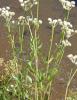
Numerous tiny white flowers are arranged in compound umbels composed of many smaller umbellets. The inflorescences create the flat or gently domed lace-like appearance that makes the species valuable as bouquet filler.
After flowering, the plant develops small, laterally compressed, ribbed dry fruits. Each fruit ultimately separates into two mericarps, each carrying one seed. Mature fruiting umbels may turn tan or brown and can scatter large numbers of phototoxic units into soil, harvested forage, poultry yards, and floral waste.
False Queen Anne’s Lace Versus Wild Carrot
True Queen Anne’s Lace or Wild Carrot is Daucus carota. It commonly has noticeably hairy stems, very finely divided foliage, a carrot-like odor when crushed, and flower heads that may curl inward into a bird’s-nest form during fruiting. Some umbels have a dark central floret, but its absence does not exclude Wild Carrot.
Ammi majus is generally taller, smoother, and more open, with large lace-like umbels that tend to remain flat or gently domed. Cut stems, immature plants, cultivated varieties, and photographs without leaves or fruit may still be difficult to distinguish.
Poison Hemlock and Water Hemlock
Poison Hemlock, Conium maculatum, contains piperidine alkaloids capable of producing salivation, tremors, weakness, ascending paralysis, respiratory failure, and death. Its smooth hollow stems commonly have irregular reddish-purple blotches, and the foliage has a markedly different finely divided appearance and odor.
Water Hemlock, species of Cicuta, is a violent convulsant plant associated especially with wet habitats. Its roots and root crown can contain concentrated cicutoxin. Early salivation, violent seizures, paddling, jaw clamping, respiratory distress, and rapid collapse are not a reason to wait for sunlight-dependent skin lesions.
When a white-flowered Apiaceae cannot be identified confidently, the exposure should be treated as potentially acute and life-threatening. Do not taste the plant or rely on a common name supplied by a seller.
Ammi visnaga and Other Bishop’s Weeds
Ammi visnaga, known as Khella or Toothpick Plant, is related but chemically and toxicologically distinct. It is better known for furanochromones such as khellin and visnagin. A controlled gosling comparison found that an equal weight of A. majus seed caused photosensitization while A. visnaga seed did not produce the same response under the study conditions.
Bishop’s Weed may also mean Goutweed, Aegopodium podagraria, or Ajwain, Trachyspermum ammi. The word ammi in a product or common name does not prove that the material is Ammi majus.
Where Animals Encounter It
Cattle, sheep, goats, and horses may encounter False Queen Anne’s Lace in pasture margins, crop stubble, flower-farm waste, hay fields, drainage areas, fence lines, or harvested forage. Sheep and cattle have the strongest direct mammalian evidence.
Poultry may consume mature seed scattered from cultivated plants, seed-cleaning waste, garden trimmings, contaminated grain, or dried flower heads. Ducks and geese kept outdoors are especially important because their beaks, eyelids, legs, and foot webs receive substantial sunlight.
Dogs and cats most often encounter the plant in gardens, bouquets, florist work areas, roadside vegetation, or dropped flower-farm material. A pet may chew stems indoors and then move to a sunny window, patio, yard, or vehicle where UVA activates absorbed compounds.
Cut Flowers and Occupational Transfer
Ammi majus is widely used as a white filler flower in wedding arrangements, cottage bouquets, centerpieces, and commercial cut-flower production. Fresh stems may release sap during cutting, stripping, bunching, transport, or disposal.
Sap can remain on gloves, clippers, counters, sleeves, towels, floors, buckets, vehicle surfaces, collars, halters, and fur. A pet or handler may be injured after contact even when the original plant is no longer nearby.
Cut stems and fallen flowers should be collected immediately. Mature fruiting heads and seed should never be placed in poultry feed, livestock forage, bedding, accessible compost, or household dried arrangements where loose fruits can fall within reach.
Seed and Forage Risk
Mature fruit and seed are the highest-priority materials because they contain a complex, directly demonstrated furanocoumarin mixture and have repeatedly produced disease in controlled animal studies.
Seed-bearing plants can enter hay, straw, crop residue, or bedding. Grinding or pelleting may prevent animals from selecting the plant out of the ration and can distribute the phototoxic material throughout a batch.
Drying does not establish safety. Mature psoralens can remain in stored botanical material, and no validated curing, ensiling, or weathering process has been shown to make contaminated forage safe for animals.
Historical and Pharmaceutical Use
Preparations derived from Ammi majus have a long history in the treatment of vitiligo. Xanthotoxin became methoxsalen, a pharmaceutical photosensitizer used with controlled UVA exposure in PUVA therapy.
This history demonstrates the plant’s mechanism rather than its safety. Medical treatment uses a known dose, planned UVA exposure, eye protection, patient selection, and clinical monitoring. Raw seed powder, homemade tincture, bouquet material, or an unknown amount consumed by an animal provides none of those controls.
The plant, its seed, and homemade extracts should never be given to an animal to alter pigmentation, treat skin disease, stimulate tanning, or reproduce human phototherapy.
Diagnosis
There is no routine veterinary test that identifies all Ammi majus furanocoumarins immediately. Diagnosis depends on accurate plant identification, exposure history, lesion distribution, the relationship between signs and light, and exclusion of hepatogenous photosensitization and other phototoxic plants.
Useful evidence includes the whole flowering or fruiting plant, leaves, lower and upper stem sections, mature umbels, seeds or mericarps, bouquet labels, seed packets, photographs of the growing site, hay samples, feed, crop residue, and material recovered from the gastrointestinal tract.
Testing may include complete blood count, serum chemistry, bilirubin, bile acids, gamma-glutamyltransferase, alkaline phosphatase, kidney values, hydration markers, electrolytes, urinalysis, and assessment for secondary infection. Specialized chromatography can identify individual furanocoumarins in plant material, but it is not routinely available as an emergency clinical test.
Ocular examination may require fluorescein staining, magnification, tonometry, examination of the anterior chamber, pupillary testing, and assessment of the retina when vision or pupil function is abnormal. Skin biopsy can help characterize phototoxic dermatitis when the diagnosis remains uncertain.
Prevention
Control the plant before fruits mature in animal areas, hay fields, poultry runs, and crop margins. Maintain adequate safe forage so grazing animals are less likely to consume unfamiliar plants.
Do not place seed-cleaning waste, flower-farm debris, bouquets, or dried umbels in paddocks, poultry yards, rabbit enclosures, kennels, barns, or accessible compost. Inspect purchased hay and bedding when unexplained photosensitization affects several animals.
Flower growers and gardeners should wear gloves and protective clothing, wash exposed skin promptly, clean tools and work surfaces, and avoid sunlight after sap contact. Pets should be excluded from the work area until all fragments and contaminated rinse water have been removed.
Immediate Steps After False Queen Anne’s Lace Exposure
- Stop further ingestion or contact. Remove the animal from the plants, mature fruit, seed, bouquets, clippings, hay, bedding, crop residue, spilled seed, and contaminated equipment.
- Begin complete light exclusion immediately. Move the animal into a darkened, well-ventilated building, stall, room, carrier, or trailer. Do not wait for redness, blistering, or squinting to begin.
- Block window and reflected UVA. Ordinary indoor shade, an open-sided shelter, a tree, or a bright room beside a window may not provide adequate protection. Cover windows and bright openings while maintaining airflow and safe temperature.
- Identify the plant. Determine whether the exposure involved Ammi majus, Wild Carrot, Ammi visnaga, Poison Hemlock, Water Hemlock, Giant Hogweed, Wild Parsnip, or another white-flowered Apiaceae.
- Determine the route and material. Record whether the animal swallowed leaves, flowers, developing fruits, mature seeds, hay, or an extract, or whether sap contacted the skin, fur, wool, feathers, or eyes.
- Remove only loose material visible at the front of the mouth. Do not force the jaws open or reach blindly into the throat.
- Preserve evidence. Save the complete plant, seed packet, bouquet label, hay, feed, crop residue, photographs, and any recovered fruits or seeds in a sealed container.
- Contact a veterinarian promptly. Seed ingestion, substantial or repeated exposure, livestock or poultry involvement, direct eye contact, uncertain identification, or any developing skin or ocular sign warrants professional guidance.
Do Not Attempt Unsupervised Home Treatment
- Do not induce vomiting. Do not use hydrogen peroxide, salt, mustard, syrup of ipecac, detergent, dish soap, manual gagging, or fingers in the throat.
- Do not force food or water. A weak, painful, vomiting, neurologically abnormal, or poorly swallowing animal may aspirate material into the lungs.
- Do not administer activated charcoal yourself. Charcoal may be aspirated and does not reverse furanocoumarin already absorbed into tissue.
- Do not apply human sunscreen automatically. Some ingredients are inappropriate when licked, and sunscreen cannot substitute for complete light exclusion. It should not be applied to eyes, open wounds, blistered tissue, or necrotic skin.
- Do not apply peroxide, alcohol, bleach, solvents, essential oils, or caustic disinfectants. These products increase tissue injury.
- Do not give owner-selected corticosteroids, antihistamines, antibiotics, pain medication, or eye drops. These products do not neutralize psoralens and may worsen infection, corneal ulceration, gastrointestinal injury, or wound healing.
- Do not assume one night indoors is sufficient. The photosensitive period depends on dose, repetition, absorption, species, and tissue injury and cannot be determined from a fixed household timetable.
Skin, Fur, Wool, and Feather Decontamination
Wear gloves, long sleeves, and eye protection when handling a contaminated animal or plant. Furanocoumarin-containing sap can injure human skin after subsequent light exposure.
Prevent grooming and wash exposed skin, paws, fur, wool, or feathers with mild liquid soap and generous cool or lukewarm water. Rinse thoroughly without aggressive scrubbing. Scrubbing inflamed skin can drive contamination into damaged tissue and rupture early blisters.
Washing reduces surface contamination but cannot remove compound already absorbed into tissue. Keep all washed areas protected from UVA afterward. Clean collars, harnesses, halters, fly masks, blankets, brushes, clippers, trailers, tools, floors, and bedding that may carry sap or seed dust.
Eye Exposure
Begin irrigation immediately with sterile saline or clean lukewarm water. Flush continuously and gently for at least 15–20 minutes, directing runoff away from the opposite eye and mouth.
Do not rub the eye, pry the eyelids forcefully apart, or use soap, peroxide, milk, contact-lens cleaner, sunscreen, or leftover ophthalmic medication. Move the animal into darkness and prevent scratching or rubbing when this can be done safely.
Photophobia, continued squinting, tearing, eyelid swelling, cloudiness, abnormal pupil size, discharge, inability to open the eye, or apparent visual impairment requires urgent veterinary examination. Ocular disease may progress after surface plant material has been flushed away.
Safe Transport Without Further Light Activation
Transport should minimize direct and reflected daylight while preserving ventilation and temperature control. Darken vehicle or trailer windows, use a covered carrier, and plan the shortest safe route.
Do not wrap an animal so tightly that breathing or cooling is impaired. Blankets, fly masks, and protective clothing must not rub blistered tissue or obstruct vision, nostrils, or airflow.
Night transport may reduce exposure when medically safe, but urgent respiratory, ocular, neurologic, or systemic signs should not be left untreated merely to wait for darkness.
Professional Gastrointestinal Decontamination
A veterinarian may consider professional emesis in an appropriate dog or cat after a recent substantial ingestion when the patient is fully alert, neurologically normal, breathing normally, not already vomiting, and able to protect the airway.
Emesis is inappropriate after weakness, collapse, tremors, seizures, respiratory abnormality, impaired swallowing, repeated vomiting, sedation, or clinically important skin or eye disease develops. Horses, cattle, sheep, goats, rabbits, guinea pigs, and other species that cannot vomit safely should never receive a home emetic.
Activated charcoal may be considered professionally after a substantial recent ingestion when aspiration risk is controlled. Exact-species evidence defining its benefit is limited, and charcoal cannot reverse tissue injury already initiated by absorbed furanocoumarins.
Large-animal veterinarians may consider rumen or gastric management after concentrated seed ingestion. The benefit must be weighed against stress, aspiration, procedural delay, and additional light exposure.
Veterinary Examination and Diagnostic Priorities
The veterinarian will assess skin pigmentation and lesion distribution, ocular pain and vision, hydration, body temperature, ability to eat and drink, wound contamination, fly strike, and evidence of another toxin.
Blood testing may include complete blood count, serum chemistry, bilirubin, bile acids, gamma-glutamyltransferase, alkaline phosphatase, albumin, kidney values, electrolytes, and markers of inflammation. These tests help distinguish primary phototoxicity from hepatogenous photosensitization and identify dehydration, infection, or organ dysfunction.
Severe weakness, tremors, paralysis, seizures, respiratory distress, or rapid collapse is not the expected defining presentation of uncomplicated Ammi majus photosensitization. Poison Hemlock, Water Hemlock, pesticides, cyanogenic plants, true nitrate-contaminated forage, and other acute toxins must be investigated.
Pain and Inflammation Control
Veterinary analgesia is often necessary because phototoxic dermatitis can be profoundly painful. Medication selection must account for species, hydration, gastrointestinal status, kidney function, pregnancy, and the extent of tissue injury.
Anti-inflammatory medication may be useful during selected phases. Corticosteroids are not a furanocoumarin antidote and require caution when infection, delayed wound healing, gastrointestinal ulceration, corneal ulceration, pregnancy, or extensive necrotic tissue is present.
Antihistamines do not neutralize psoralens or prevent DNA photoadduct formation. They may have a role when a separate allergic process or specific inflammatory component is suspected, but they cannot replace darkness, analgesia, wound care, and ocular monitoring.
Wound Care and Skin Necrosis
Damaged skin may require gentle cleansing, nonadherent dressings, moist-wound management, topical barriers selected for the species, and protection from urine, manure, dirt, and friction.
Devitalized tissue should be evaluated repeatedly. Debridement may be delayed until tissue viability is clear or performed in stages. Owners should not cut, peel, or pull away attached skin.
Antimicrobial treatment is appropriate when bacterial infection, cellulitis, fever, sepsis, or heavily contaminated necrotic tissue is present. Antibiotics are not automatically required for every erythematous lesion and do not stop photochemical injury.
Fly-strike prevention is essential in livestock and outdoor animals. Housing should exclude flies, and affected skin should be checked repeatedly for eggs or larvae. Established myiasis requires immediate removal of larvae, wound treatment, and species-appropriate parasite control.
Fluid, Nutritional, and Temperature Support
Intravenous fluids may be required when painful muzzle or eye lesions prevent drinking, extensive wounds lose fluid, fever or infection develops, or the animal becomes dehydrated.
Soft feed, protected feeders and waterers, assisted feeding, or other nutritional support may be necessary when lip, muzzle, beak, or ocular disease interferes with normal intake. Force-feeding an animal that cannot swallow safely is inappropriate.
Severely affected animals should be housed in a cool, clean, dark environment. Extensive skin damage can impair normal temperature regulation, while an enclosed building can become dangerously hot without adequate ventilation.
Veterinary Ocular Treatment
Ocular examination may include fluorescein staining, magnification, pupillary testing, tonometry, anterior-segment examination, and assessment of the retina and choroid when visual function is abnormal.
Treatment may involve ocular lubrication, pain control, cycloplegic medication, topical antimicrobial therapy when epithelial defects create infection risk, and other medication chosen after the cornea has been examined.
Topical corticosteroids should not be administered blindly. They may worsen an untreated corneal ulcer, delay epithelial healing, or increase infection risk. Persistent mydriasis, eyelid adhesion, retinal injury, or corneal scarring may require prolonged ophthalmic care.
Cattle, Sheep, Goats, and Horses
Move the entire exposed group into dark housing and remove the shared pasture, hay, crop residue, or seed source. Examine apparently normal animals because lesion severity can differ with pigmentation, individual intake, and light exposure.
Provide feed and water inside the protected enclosure. Animals should not be forced into bright yards for repeated examination, milking, weighing, or photography while active photosensitivity is suspected.
White facial markings, udders, teats, vulvas, coronary bands, and distal limbs require repeated inspection. Protective masks or coverings may be useful under veterinary direction, but they must not trap heat, moisture, flies, or contaminated exudate against damaged skin.
Poultry and Waterfowl
Move birds into a dark, ventilated building and remove all seed, plant waste, litter, and contaminated feed. Examine every bird sharing the exposure.
Pay particular attention to the beak, eyelids, eyes, comb, wattles, legs, feet, and foot webs. Young birds may develop permanent deformity as scar tissue contracts during growth.
Place food and water where visually impaired birds can locate them without competition. Birds with severe beak deformity, eyelid adhesion, mydriasis, retinal damage, or painful foot lesions may require prolonged supportive care and individual welfare assessment.
Prognosis and Duration of Light Restriction
The prognosis is best when exposure is recognized before prolonged UVA activation. Mild erythema and edema may improve over several days, but ulceration, necrosis, corneal disease, and chronic poultry deformity can require weeks or months of care.
No fixed number of dark-housing hours is safe for every exposure. Experimental xanthotoxin-disposition work found rapid elimination of much of a single administered dose in hens and a goat, but plant ingestion may be repeated, compound mixtures differ, tissue-bound photoproducts remain active, and clinical lesions can continue evolving after circulating material declines.
Return to sunlight should be gradual and veterinarian-directed. The animal should remain comfortable under increasing controlled light, without new erythema, edema, photophobia, or lesion progression.
Frequently Asked Questions About False Queen Anne’s Lace and Animal Poisoning
How long must an exposed animal be kept out of sunlight?
No universal interval has been established. The duration depends on the amount and frequency of exposure, the furanocoumarin mixture, species, absorption, pigmentation, and whether lesions have already developed. A radiolabeled-xanthotoxin study found that hens and a goat eliminated most of a single dose relatively quickly, but that does not establish that every plant-exposed animal is safe after 24 or 48 hours. Repeated ingestion, continued absorption from the gastrointestinal tract, tissue photoproducts, and evolving wounds can extend risk. Light should be reintroduced gradually under veterinary direction only after the animal remains comfortable and no new lesions appear.
Is a bright room or barn with windows dark enough?
Not necessarily. Window glass blocks much of UVB but can transmit a meaningful portion of UVA, the wavelength range that activates psoralens. Reflected daylight entering through doors, translucent roof panels, open sides, and pale surfaces may also continue activation. Affected animals require a substantially darkened but well-ventilated enclosure. Windows and bright openings should be covered without creating dangerous heat or poor airflow.
Why can skin damage worsen after the plant has been removed?
Removing the source stops additional exposure but does not remove furanocoumarin already absorbed into the blood or deposited in tissue. UVA can continue activating that material, and cells already carrying DNA cross-links and membrane damage may progress to apoptosis or necrosis over time. Inflammation, vascular leakage, secondary infection, and wound contraction can also continue after the original photochemical event.
Can washing the skin completely prevent photosensitization?
Prompt washing can substantially reduce unabsorbed sap and is especially important after direct contact. It cannot extract material that has already penetrated the skin or entered the circulation after ingestion. Washed skin must still be protected from UVA, and the animal must be monitored for delayed pain, redness, edema, blistering, and ocular signs.
Can a black or dark-coated animal still be injured?
Yes. Dark pigment and dense hair reduce light penetration but do not protect the eyes, eyelids, nasal planum, lips, vulva, teats, foot pads, thin-haired areas, clipped skin, wounds, or directly contaminated sites. A dark animal may have less widespread skin disease while still developing severe ocular injury or sharply localized contact-pattern dermatitis.
Why do some sources call the toxic material seed while others call it fruit?
The small unit commonly planted or found in feed is botanically a dry fruit. At maturity it divides into two mericarps, and each mericarp contains one seed. Veterinary and agricultural papers frequently use “seed” for the complete dried unit. The terminology difference does not make one form safer; intact fruits, separated mericarps, and their enclosed seeds should all be treated as phototoxic material.
Is Ammi visnaga equally photosensitizing?
It should not be assumed to be equivalent. In a controlled gosling comparison, the same weight of Ammi majus seed caused photosensitization while Ammi visnaga seed did not produce the same clinical response. A. visnaga contains a different dominant chemical profile, including khellin and visnagin. Both species require accurate identification, but evidence from one should not be copied directly to the other.
Why are liver tests needed when Ammi majus causes primary photosensitization?
Primary photosensitization does not require liver injury, but the skin pattern can resemble hepatogenous photosensitization caused by phylloerythrin retention. Liver and bile-duct testing helps distinguish the mechanisms and may reveal another plant exposure, facial eczema, biliary obstruction, or concurrent disease. Normal liver-associated results support a primary process but do not prove that Ammi majus was the source.
Does False Queen Anne’s Lace cause nitrate poisoning?
Nitrate poisoning has been repeated in secondary weed lists, but exact-species evidence is weak. Research showing nitrate uptake, nitrate-reductase activity, or increased plant nitrogen is not proof that animals developed methemoglobinemia from toxic Ammi majus forage. Rapid breathing, chocolate-brown blood, blue-gray mucous membranes, weakness, or sudden deaths still requires immediate nitrate testing and treatment, but other forage plants, fertilizer contamination, water, and crop residues must be investigated rather than assuming that Ammi caused the syndrome.
Do calcium oxalate crystals explain the poisoning?
No. Microscopic rosette-shaped calcium oxalate deposits have been described in the fruit, but this does not establish forcefully discharged raphides, severe aroid-type oral injury, systemic hypocalcemia, or oxalate nephrosis. Direct chemistry and controlled animal studies identify furanocoumarin phototoxicity as the defining hazard. Immediate severe oral burning should prompt confirmation of the plant and investigation for another irritant species or contaminant.
Can poultry suffer permanent injury after the skin appears to heal?
Yes. Scar contraction during growth can leave geese and ducks with shortened or deformed upper beaks, thickened and contracted foot webs, or eyelids scarred partly or completely together. Mydriasis may persist after damage to the iris sphincter, and pigmentary retinal disease may cause lasting visual impairment. These chronic effects are why early darkness and ocular protection matter even when acute mortality is low.
Can dried bouquet stems and seed heads still cause poisoning?
Yes. Drying does not prove that xanthotoxin, bergapten, isopimpinellin, or the other seed psoralens have disappeared. Dried fruiting heads can shed phototoxic material onto floors, tables, feed, and bedding. Florist preservatives, wire, picks, glue, paint, or other flowers may create additional hazards, so the entire arrangement should be preserved for identification after an exposure.
What should be done when Poison Hemlock or Water Hemlock cannot be excluded?
Treat the exposure as an acute emergency. Do not wait for a delayed photosensitive rash. Poison Hemlock can cause weakness, tremors, paralysis, and respiratory failure, while Water Hemlock can cause violent seizures and rapid death. Keep the animal quiet, prevent further ingestion, preserve the complete plant, avoid owner-administered emetics or medication, and obtain immediate veterinary care.