PAWS Pet Poison Plant Guide
Is Bishop’s Weed Poisonous to Dogs, Cats, Horses, and Livestock?
Yes, Bishop’s weed, Ammi majus, is poisonous to dogs, cats, horses, livestock, rabbits, poultry, and other animals. Its furanocoumarins can make the skin and eyes abnormally sensitive to sunlight after the plant is eaten or its sap contacts exposed tissue. Injury may include painful redness, swelling, blistering, crusting, ulceration, skin sloughing, squinting, tearing, corneal cloudiness, and impaired vision. Fruits and seeds are especially rich in photosensitizing compounds, but all parts should remain inaccessible.
About this guide: This page provides general pet-poisoning information and cannot diagnose or treat an individual animal. For any suspected exposure, contact a veterinarian or animal poison-control service immediately. Do not induce vomiting, give medication, or attempt home decontamination unless directed by a veterinary professional.
Bishop’s Weed
Ammi majus L.
Apiaceae
Umbelliferae is the older alternative family name encountered in historical botanical and veterinary literature.
Bishop’s Weed, Bishop’s Flower, False Bishop’s Weed, False Queen Anne’s Lace, Greater Ammi, Large Bullwort, Bullwort, Laceflower, Lace Flower, Lady’s Lace, White Dill, Greater Bishop’s Weed, Ammi, Ammi majus
“Queen Anne’s Lace” properly refers most often to Daucus carota and should not be treated as an exact name for Ammi majus. “Bishop’s Weed” is also used for unrelated plants, including Aegopodium podagraria, Trachyspermum ammi, and other members of the Apiaceae.
Linear Furanocoumarins Are the Principal Phototoxins
The principal toxins in Bishop’s weed are linear furanocoumarins, also called psoralens. These compounds include xanthotoxin, bergapten, isopimpinellin, isoimperatorin, imperatorin, and related molecules. Xanthotoxin is also known as 8-methoxypsoralen or methoxsalen, while bergapten is 5-methoxypsoralen.
Furanocoumarins are unusual because the plant compound and ultraviolet radiation must act together to produce the characteristic injury. A photosensitizing compound may enter the animal after ingestion or remain on tissue after direct sap contact. When the exposed skin or eye then receives sufficient ultraviolet A radiation, the compound becomes photochemically activated and damages the tissue.
This is not an ordinary caustic burn produced immediately by plant juice alone. An animal may eat the plant or acquire sap on the skin and remain outwardly normal while indoors, in darkness, or before enough ultraviolet exposure has occurred. Painful inflammation, blistering, ulceration, and ocular injury can appear later when the animal enters sunlight.
The Texas Seed Study Identified Twelve Psoralens
G. Wayne Ivie published “Linear Furocoumarins (Psoralens) from the Seed of Texas Ammi majus L. (Bishop’s Weed)” in 1978. Twelve linear furanocoumarins were isolated from ripe seeds collected from Texas plants.
The confirmed compounds included xanthotoxin, bergapten, isopimpinellin, and isoimperatorin. The study also identified additional known psoralens not previously recovered from the species and two compounds that had apparently not previously been isolated from any plant source.
Most of the major seed psoralens demonstrated potent photosensitizing activity in chick-skin bioassays. This work directly supports the conclusion that ripe Bishop’s-weed seeds contain a mixture of active phototoxic compounds rather than one uniform toxin.
A modern analysis of Ammi majus fruits independently isolated and quantified isopimpinellin. The fruit and seed evidence is therefore especially strong, although the exact concentration varies with plant material, population, maturity, extraction method, and analytical conditions.
Primary Photosensitization
Bishop’s weed causes primary photosensitization. The plant itself supplies the photodynamic compounds, which are absorbed or deposited in the tissue before light activation. Pre-existing liver failure is not required.
This differs from hepatogenous or secondary photosensitization. In that syndrome, liver or bile-duct dysfunction prevents normal elimination of phylloerythrin, a photodynamic chlorophyll-breakdown product generated in the digestive tract. Phylloerythrin then accumulates in the bloodstream and skin.
The outward skin lesions can look similar in both forms. For that reason, a veterinarian may still evaluate liver enzymes, bilirubin, bile acids, feed history, other poisonous plants, medications, and systemic disease rather than assuming every photosensitive animal has uncomplicated primary Ammi majus poisoning.
How Ultraviolet Light Activates the Toxins
Linear furanocoumarins absorb ultraviolet A energy. After photoactivation, the molecules can interact with cellular DNA and form covalent photoadducts. Some can produce interstrand cross-links that interfere with DNA replication and transcription.
Photoactivation also damages proteins, cell membranes, keratinocytes, vascular structures, and other cellular targets. Reactive oxygen species and inflammatory mediators contribute to edema, pain, erythema, blistering, vascular leakage, epidermal necrosis, and delayed tissue sloughing.
The reaction may continue to evolve after the animal has been removed from direct sunlight because cellular damage and inflammation already initiated in the tissue do not stop instantly. Repeated ultraviolet exposure during the photosensitive period can deepen existing injury or create new lesions.
Standard window glass blocks much ultraviolet B but can transmit a portion of ultraviolet A. An exposed animal should therefore be housed away from bright windows and glass doors rather than merely moved to a sunny indoor room.
Cattle and Sheep Feeding Studies
J. W. Dollahite, R. L. Younger, and G. O. Hoffman experimentally produced photosensitization in cattle and sheep by feeding Ammi majus. They also observed phototoxic injury in people who had dermal contact with the plant followed by sunlight exposure, confirming that both ingestion and direct contamination can be clinically important.
D. A. Witzel, J. W. Dollahite, and L. P. Jones separately fed finely ground Bishop’s-weed seed to sheep at experimental exposure levels of 1, 2, 4, and 8 grams per kilogram of body weight. Repeated exposure at 2 and 4 grams per kilogram produced comparable photosensitization within approximately 72 to 96 hours.
These experimental amounts should not be converted into a household pet threshold. Ground seed delivered experimentally to sheep differs from a dog tasting a flower, a cat contacting sap, a horse grazing whole plants, or poultry consuming seeds mixed into feed. The studies establish hazard and dose responsiveness, not a universally applicable safe amount.
Confirmed Field Disease in Cattle
A later cattle outbreak in Argentina provided direct field evidence of primary photosensitization associated with voluntary consumption of Ammi majus. Affected cattle developed photophobia, excessive tearing, bilateral corneal opacity, and dermatitis involving the muzzle, vulva, udder, and teats.
The investigators excluded secondary photosensitization associated with liver injury and identified consumption of Bishop’s weed as the cause. This report is especially important because it documents the distribution of lesions expected in naturally exposed grazing animals rather than only under controlled experimental conditions.
Poultry Studies Demonstrated the Sunlight Requirement
Experimental work in geese, ducks, ducklings, chickens, and turkeys established that ingestion alone did not produce the same visible disease when ultraviolet exposure was prevented. Goslings fed Ammi majus seed and exposed to sunlight developed photosensitization, while comparable birds protected from sunlight did not develop the clinical syndrome.
A comparative experiment also distinguished Ammi majus from the related toothpick plant, Ammi visnaga. Goslings fed A. majus seed developed photosensitization after sunlight exposure, while birds fed equal weights of A. visnaga stems, leaves, and flowers did not develop the same clinical disease.
Young chickens showed no visible effect when seeds formed 1.25 percent of the experimental diet, while a 3 percent seed diet produced mild photosensitization within approximately six to eight days. Ducks and turkeys differed in susceptibility. These species differences show why one poultry exposure level cannot be assigned to every bird.
Acute and Chronic Injury in Ducks and Geese
M. N. Egyed, A. Shlosberg, A. Eilat, Mertyn Malkinson, and colleagues documented acute and chronic manifestations of Ammi majus-induced photosensitization in ducks. Acute lesions were inflammatory, while chronic disease included severe deformation of the beak and foot webs, dilated or abnormally positioned pupils, and lasting ocular damage.
In a naturally affected goose flock, 64 of 133 birds showed chronic abnormalities seven weeks after the acute disease began. All affected birds had stunting of the upper beak and cicatrization with thickening of the foot webs. Ocular involvement occurred in most affected birds and included blepharoconjunctivitis, keratitis, adhesions of the eyelids or conjunctiva, eyelid distortion, and mydriasis.
These findings demonstrate that the syndrome is not limited to a temporary skin rash. Severe or repeated phototoxic injury can alter growing structures, contract healing tissue, deform the beak or feet, and permanently impair ocular anatomy and vision.
Direct Ocular Toxicity and Long-Term Retinal Injury
Experimental studies specifically examined the eyes of geese and ducks photosensitized by ingestion of Bishop’s-weed seeds. Mydriasis was a characteristic finding. Histologic examination found vacuolization and atrophy of the iris sphincter muscle, helping explain persistent pupil dilation.
Ducklings force-fed Ammi majus seeds for five days and exposed to sunlight developed acute and chronic retinal and choroidal lesions. Early findings included vacuolization of retinal ganglion cells. Birds examined months later showed pigmentary retinopathy, hyperplasia of the retinal pigment epithelium, vascular congestion, dilation, and fluid leakage in the choroid.
Separate experimental work documented eye lesions in ducklings fed Bishop’s-weed seed. These studies support urgent examination of squinting, tearing, corneal haze, apparent blindness, abnormal pupils, or persistent light avoidance rather than treating the eye signs as secondary to skin discomfort alone.
Ingestion Versus Direct Sap Contact
Ingestion can produce systemic photosensitization because absorbed furanocoumarins circulate to light-exposed tissues. Lesions may therefore appear on several unpigmented or sparsely covered areas even when plant sap never touched those sites directly.
Direct contact can create sharply localized phytophotodermatitis. Sap or crushed plant material deposited on the muzzle, paws, lower abdomen, udder, teats, genital skin, or another exposed area may later produce linear streaks, handprint-like patches, splash patterns, or irregular zones of inflammation after ultraviolet exposure.
A localized pattern does not prove that the animal failed to ingest the plant, and generalized disease does not rule out additional direct-contact injury. An animal that chewed, rolled in, or walked through the plant may experience both routes simultaneously.
Why White and Sparsely Covered Skin Is Most Vulnerable
Melanin, hair, wool, and feathers reduce penetration of ultraviolet radiation. White, lightly pigmented, recently clipped, thinly haired, or unfeathered tissues receive less protection and are therefore injured most severely.
Commonly affected sites include white facial markings, eyelids, nostrils, lips, ear margins, the sparsely haired abdomen, lower limbs, udder, teats, vulva, prepuce, and other exposed mucocutaneous junctions.
Dark pigmentation does not provide complete immunity. Intense ultraviolet exposure, a large phototoxin burden, sparse hair, an exposed eye surface, or direct sap contamination may still injure pigmented animals.
Fruits, Seeds, and the Rest of the Plant
Ripe fruits and seeds contain the best-characterized and most concentrated mixture of linear furanocoumarins. Seed ingestion produced disease in sheep and poultry experiments, and ripe seed chemistry has been analyzed directly.
Leaves, stems, flowers, developing seed heads, and sap should also be treated as hazardous. Whole-plant feeding produced photosensitization in cattle and sheep, and dermal contact followed by sunlight caused human phototoxic injury in experimental and clinical observations.
No safe leaf count, stem weight, flower number, fruit quantity, seed number, or amount per kilogram has been established for dogs, cats, horses, rabbits, or individual livestock species. Exposure assessment must consider the plant part, amount, animal species, pigmentation, coat coverage, and ultraviolet exposure.
Dried Plants, Hay, Bouquets, and Stored Seeds
Dried fruits, seed heads, seeds, hay contamination, cut flowers, discarded arrangements, and harvested plant material should not be considered safe. The principal furanocoumarins are sufficiently persistent to be isolated from dried fruits and seeds used in chemical and pharmacological research.
Drying may change moisture, enzyme activity, texture, and palatability, but it does not reliably eliminate the phototoxic molecules. Seed-contaminated poultry feed and dried plant material remain meaningful exposure concerns.
The Nitrate Claim Is Not Established
No adequate species-specific research was located establishing Bishop’s weed as a clinically important nitrate accumulator or documenting nitrate poisoning caused by authenticated Ammi majus. Nitrate should therefore not be presented as a second recognized toxin of this plant.
An animal with rapid breathing, weakness, tremors, chocolate-brown mucous membranes, brown blood, collapse, or sudden death requires immediate investigation for nitrate-contaminated forage, fertilizer, another plant, smoke or oxidant exposure, and other causes of methemoglobinemia. Those signs should not be attributed automatically to Bishop’s weed.
A Delayed Reaction After Ingestion or Contact
An animal may appear completely normal immediately after eating Bishop’s weed or acquiring sap on the coat. The defining injury develops after phototoxic compounds in or on the tissue receive sufficient ultraviolet A exposure.
The interval between plant exposure and visible disease depends on the amount absorbed, plant part, route of exposure, pigmentation, hair or feather coverage, ultraviolet intensity, time spent in sunlight, and individual susceptibility. Signs may begin within hours of light exposure or become much more apparent over the following day.
The absence of redness at the time the plant is discovered does not make continued outdoor exposure safe. Moving the animal indoors or into genuinely complete shade is an immediate protective measure rather than something reserved for animals that have already blistered.
Early Pain, Restlessness, and Light Avoidance
Early photosensitization often causes intense skin sensitivity before obvious tissue destruction is visible. Animals may become restless, agitated, unwilling to be touched, or unusually distressed when exposed to sunlight.
Dogs may rub or paw at the muzzle, seek furniture or dark rooms, resist going outdoors, or react painfully when light reaches affected skin. Cats may hide, overgroom, shake the head, rub the face, or avoid bright areas.
Grazing animals may seek trees, sheds, fence shadows, or buildings. They may stamp, kick at the abdomen, rub against posts, shake the head, or become difficult to move into sunlight. Poultry may crowd beneath shelters or refuse to leave shaded areas.
Progressive Skin Injury
Exposed skin may initially become warm, red, painful, and swollen. Edema can be especially marked around the eyelids, muzzle, lips, ears, udder, teats, vulva, prepuce, and lower limbs.
As the injury progresses, vesicles or larger blisters may form. Serum can seep through damaged tissue, mat the hair or feathers, and dry into thick crusts. Fissures, erosions, ulcers, and sharply demarcated necrotic areas may follow.
Severely damaged epidermis can separate and slough, leaving moist, raw wounds. Sloughing may not become fully apparent until days after the original sunlight exposure because the injured tissue must first lose viability and detach.
Lesions are often most dramatic on white or unpigmented skin while adjacent dark skin is relatively spared. This boundary can be diagnostically useful but is not present in every animal, particularly when direct sap contact creates an irregular localized pattern.
Commonly Affected Body Sites
Frequently affected areas include the ear margins, eyelids, nose, muzzle, lips, white facial markings, sparsely haired abdomen, footpads, lower limbs, udder, teats, vulva, prepuce, and other exposed skin.
Recently clipped or shaved areas may be unusually vulnerable. Surgical clipping, grooming, shearing, dermatologic hair loss, wet feathers, and damaged skin can reduce normal physical protection from ultraviolet radiation.
In poultry and waterfowl, the eyelids, beak, legs, feet, and foot webs are especially exposed. Chronic contraction of injured tissue can shorten, thicken, or deform these structures.
Ocular Pain and Vision-Threatening Injury
Eye involvement is a veterinary priority. Early signs include squinting, forceful eyelid closure, light avoidance, excessive tearing, eyelid swelling, conjunctival redness, and rubbing or pawing at the face.
Corneal involvement may produce blue-gray haze, white opacity, surface roughness, ulceration, abnormal blood-vessel growth, or apparent vision loss. Uveitis may cause a painful eye, altered pupil size, redness around the iris, and reduced vision.
Experimental bird studies documented mydriasis, displacement or distortion of the pupil, keratitis, adhesions of ocular tissues, retinal injury, pigmentary retinopathy, and chronic abnormalities of the retinal pigment epithelium and choroid.
An animal that bumps into objects, fails to track movement, holds an eye closed, develops corneal cloudiness, or has an abnormally dilated or irregular pupil requires urgent examination. Ocular injury can progress even when the skin lesions appear limited.
Localized Phytophotodermatitis After Sap Contact
Dogs and cats may develop localized lesions where fresh sap or crushed plant material contacted the body. The muzzle, paws, lower legs, abdomen, and inner thighs are common contact sites.
The pattern may be linear, streaked, splashed, or irregular rather than distributed symmetrically across every light-colored area. A dog walking through cut stems may develop lesions on the paws and ventral abdomen, while an animal chewing the plant may develop a concentrated reaction around the lips and muzzle.
Localized contact injury can coexist with systemic photosensitization after ingestion. The presence of one pattern does not exclude the other.
Dogs and Cats
Published controlled poisoning studies are concentrated in cattle, sheep, and poultry. A dependable canine or feline toxic dose has not been established. The photochemical mechanism nevertheless applies to mammalian skin and eyes, and direct human contact cases confirm that Ammi majus sap and fruit material can cause severe phototoxic dermatitis after sunlight exposure.
Dogs with white facial markings, thin coats, sparsely haired abdomens, recently clipped fur, or prolonged outdoor exposure may develop more extensive visible lesions. Cats with white ears, noses, eyelids, or facial skin may also be vulnerable.
Food refusal may result from painful lips, oral contact, ocular pain, fever, or general distress. Continued anorexia in a cat deserves prompt attention because prolonged failure to eat can create serious secondary metabolic complications.
Cattle, Sheep, Goats, and Horses
Cattle and sheep have developed primary photosensitization experimentally after eating Ammi majus. Naturally affected cattle have shown photophobia, tearing, bilateral corneal opacity, and dermatitis of the muzzle, vulva, udder, and teats.
Grazing animals may show restlessness, rubbing, head shaking, reduced grazing, reluctance to move through sunlight, weight loss, dehydration, and declining body condition. Teat injury may interfere with nursing or milking and can predispose the animal to mastitis.
Severe lesions around the vulva or prepuce can interfere with urination or breeding. Painful lower-limb lesions may cause stiffness, lameness, or reluctance to walk.
Direct horse-specific Bishop’s-weed case evidence is limited. Horses should nevertheless be protected because the established phototoxic compounds and primary photosensitization mechanism are relevant across species. White facial markings, pink muzzle skin, clipped areas, and unpigmented lower limbs would be expected to carry the greatest visible risk.
Geese, Ducks, Chickens, Turkeys, and Other Birds
Birds have some of the strongest species-specific evidence. Acute disease may include redness, swelling, inflammation of the eyelids, exudation, photophobia, foot pain, reluctance to walk, reduced feeding, and ocular injury.
Chronic disease in geese and ducks has produced stunting and deformation of the upper beak, thickening and contraction of the foot webs, keratitis, adhesions involving the eyelids and conjunctiva, abnormal pupil size or position, and lasting visual impairment.
Young chickens developed mild clinical photosensitization after consuming a higher experimental dietary concentration of Bishop’s-weed seed, while lower dietary exposure did not produce visible disease during the study period. Ducks and turkeys showed different susceptibility, demonstrating that risk cannot be predicted solely from body weight.
Pet birds should not be offered Bishop’s weed, its fruits, or its seeds as forage, enrichment, nesting material, or decorative vegetation. Seed contamination of feed is a particularly important exposure route.
Rabbits, Guinea Pigs, and Other Small Animals
Published Bishop’s-weed poisoning studies in rabbits and guinea pigs are limited, and no toxic dose has been established. These animals should not receive any part of the plant.
Rabbits and guinea pigs cannot vomit. Reduced appetite, fewer or smaller fecal pellets, a hunched posture, tooth grinding, reduced movement, eye pain, skin tenderness, or hiding from light requires prompt veterinary attention.
Pain and food refusal can contribute to gastrointestinal stasis, dehydration, altered intestinal flora, and metabolic deterioration independently of the phototoxic skin injury.
Secondary Infection, Fluid Loss, and Fly Strike
Open blisters, ulcers, necrotic tissue, and sloughed skin lose fluid and protein and create an entry point for bacteria. Warning signs of secondary infection include fever, spreading redness, progressive swelling, purulent or foul-smelling discharge, worsening pain, and declining appetite.
Outdoor animals with moist wounds are at risk of fly strike. Flies may deposit eggs in damaged tissue, and developing larvae can rapidly extend the wound, deepen tissue destruction, and cause systemic illness.
Extensive skin loss can cause dehydration, electrolyte disturbance, hypoproteinemia, heat loss, and profound debilitation. These complications can become more dangerous than the initial photochemical reaction.
Expected Course and Lasting Effects
The photosensitive state can persist after the plant is removed because absorbed compounds require time to be metabolized and eliminated. A brief return to sunlight can worsen lesions while sufficient phototoxin remains in the skin or eye.
Superficial inflammation may improve over several days, while deep ulceration, necrosis, infection, and ocular injury can require weeks of treatment. Healing skin may remain unusually sensitive during recovery.
Permanent consequences can include scarring, altered pigmentation, hair or feather loss, contraction of skin, beak or foot-web deformity, corneal scarring, abnormal pupils, retinal damage, and partial or complete vision loss.
Signs That Suggest Another or Additional Poison
Very rapid respiratory distress, chocolate-brown blood or mucous membranes, sudden generalized tremors, immediate collapse, or several unexplained deaths are not the expected uncomplicated Bishop’s-weed photosensitization pattern.
Those findings require investigation of nitrate-contaminated forage, fertilizer, cyanogenic plants, pesticides, oxidizing chemicals, smoke exposure, blue-green algae, another poisonous plant, infectious disease, or a mixed exposure.
Plant Identity and Native Range
Bishop’s weed is Ammi majus L., an annual herb in Apiaceae. Umbelliferae is the older alternative family name encountered in historical botanical, agricultural, pharmacological, and veterinary literature.
The species is native across Macaronesia and the Mediterranean region and extends eastward through western and central Asia to the Arabian Peninsula. It has been introduced into many other temperate and subtropical regions and can persist outside cultivation in disturbed ground, roadsides, field margins, drainage areas, and former ornamental plantings.
Animals may encounter it both as an intentionally grown laceflower and as a naturalized or agricultural weed. Its delicate white flower heads can make it appear harmless, but the fruits and seeds contain a particularly well-characterized mixture of potent linear furanocoumarins.
Why the Name “Bishop’s Weed” Is Unreliable
“Bishop’s weed” is applied to several unrelated plants. Ground elder is Aegopodium podagraria, while ajwain is Trachyspermum ammi. Neither should be assumed to have the same toxicology as Ammi majus.
“Queen Anne’s lace” most often refers to wild carrot, Daucus carota. Bishop’s flower, false Queen Anne’s lace, greater ammi, large bullwort, and laceflower are commonly applied to Ammi majus, but the scientific name remains necessary for reliable identification.
The related toothpick plant, Ammi visnaga, can also be confused with Bishop’s weed. Experimental gosling research found that A. majus seed caused photosensitization under sunlight while comparable A. visnaga stems, leaves, and flowers did not produce the same disease. The species should therefore not be treated as interchangeable.
Dangerous White-Umbelled Look-Alikes
Bishop’s weed belongs to a family containing numerous white-flowered plants with superficially similar compound umbels. Potential look-alikes include wild carrot, poison hemlock, water hemlock, cow parsley, wild parsnip, giant hogweed, hemlock water-dropwort, and other Apiaceae.
Some look-alikes cause rapid neurologic, respiratory, cardiovascular, or gastrointestinal poisoning rather than delayed photosensitization. Poison hemlock and water hemlock can be fatal through mechanisms entirely different from furanocoumarin phototoxicity.
Wild parsnip and giant hogweed can produce severe contact phytophotodermatitis but differ in plant size, leaf form, stem features, and exposure circumstances. An unidentified white umbel should never be diagnosed from the flower head alone.
How to Recognize Ammi majus
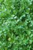
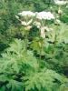
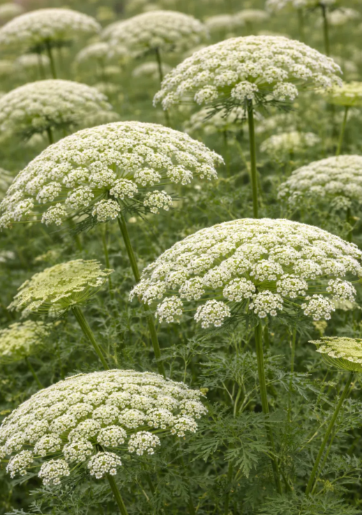
Ammi majus is an upright, branching annual that may grow from approximately one foot to more than five feet tall depending on environmental conditions. The green stems are ridged and generally hairless.
Lower leaves are pinnate or bipinnate with broader toothed segments. Upper leaves become more finely divided and commonly attach with sheathing leaf bases.
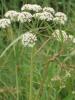
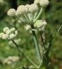
The numerous small white flowers are arranged in broad compound umbels at the ends of the stems. Individual flowers have five petals. The umbels commonly contain many slender rays and appear relatively open and lace-like.
Wild carrot may carry a small dark red or purple flower near the center of the umbel and often curls into a nest-like seed head as it matures. Those features are not completely reliable, and Bishop’s weed may vary in appearance. Photographs for identification should show the entire plant, stem, upper and lower leaves, flower heads, developing fruits, mature fruits, and base.
Where Dogs and Cats Encounter It
Dogs and cats may encounter Bishop’s weed in flower beds, cutting gardens, wildflower plantings, roadsides, field margins, vacant lots, drainage areas, bouquets, dried arrangements, discarded stems, and compost or landscape waste.
Exposure can occur when an animal chews the plant, walks through a dense stand, rolls in cut vegetation, lies on discarded stems, or brushes sap onto the muzzle, paws, legs, or abdomen.
Cut flowers brought indoors can remain hazardous. An animal may chew the stems or fruits, contact sap released during trimming, or later enter sunlight with contaminated fur.
Where Horses and Livestock Encounter It
Horses, cattle, sheep, goats, and other livestock may encounter the plant in pasture, hay, field margins, fence lines, irrigation channels, drainage areas, roadsides, feed crops, and discarded ornamental material.
Large-scale exposure is more likely when animals have limited alternative forage, consume contaminated hay, graze dense stands, or gain access to cut plants and mature seed heads.
The reproductive stage is especially important because fruits and seeds contain concentrated, chemically documented psoralens. Mature seed heads can contaminate harvested forage or remain accessible after the green portions of the plant have dried.
Poultry and Pet-Bird Exposure
Poultry may be exposed when Bishop’s-weed seeds contaminate grain or collected vegetation, when whole plants are placed in runs, or when birds forage beneath mature stands.
Geese and ducks may consume the fruits and seeds readily. Experimental studies demonstrated that seed ingestion followed by sunlight exposure can produce severe acute and chronic disease.
Dried seeds should not be added to pet-bird food, wild-bird mixtures, enrichment, or nesting material. Dry appearance does not establish that the photosensitizing compounds have been removed.
Poisonous Parts and Relative Risk
Leaves, stems, flowers, sap, developing fruits, mature fruits, and seeds should all be regarded as capable of causing exposure. Fruits and seeds present the strongest documented hazard because their psoralen chemistry has been characterized directly and seed feeding has repeatedly produced disease in animals.
Freshly cut or crushed vegetation creates an important contact hazard. Sap on the muzzle, paws, abdomen, udder, or another light-exposed site may produce localized lesions after sunlight exposure even when the amount swallowed was small.
Wilted, dried, or harvested material is not reliably safe. Furanocoumarins remain recoverable from stored fruits and seeds, and contaminated hay or dried arrangements can preserve an exposure route after the fresh plant is gone.
Why Exposure Severity Cannot Be Predicted from Plant Weight Alone
The amount of plant material matters, but ultraviolet exposure also controls the severity of visible disease. A modest ingestion followed by intense, prolonged sunlight may cause more tissue injury than a larger exposure followed by immediate light exclusion.
Skin pigmentation, coat or feather density, clipping, age, species, weather, latitude, season, cloud cover, altitude, time of day, and duration outdoors all influence the amount of ultraviolet energy reaching the photodynamic compounds.
No dependable dog, cat, horse, rabbit, poultry, or livestock toxic dose can be calculated from one leaf count or plant weight. The experimental sheep exposure levels demonstrate hazard but should not be used as owner-facing thresholds.
Primary Photosensitization Versus Liver Disease
Bishop’s weed can cause primary photosensitization directly. Nevertheless, similar lesions can result from liver disease and accumulation of phylloerythrin.
Veterinary evaluation may include liver enzymes, bilirubin, bile acids, ultrasonography, feed analysis, and investigation of hepatotoxic plants or molds. A photosensitive animal with jaundice, major liver-value abnormalities, neurologic signs associated with hepatic dysfunction, or widespread herd disease may have another or an additional cause.
Dark housing is necessary in either form while the photosensitive compound remains active, but long-term treatment and prognosis depend on the underlying mechanism.
Diagnosis
There is no routine blood test that confirms Ammi majus ingestion or provides a clinically useful furanocoumarin concentration. Diagnosis depends on plant identification, evidence of ingestion or sap contact, subsequent ultraviolet exposure, and the distribution of lesions on unpigmented or exposed tissues.
Owners should preserve a complete sample or clear photographs showing the stem, upper and lower leaves, umbels, fruits, seeds, and plant base. In livestock or poultry outbreaks, pasture plants, hay, feed, and seed contamination may require examination or laboratory analysis.
The diagnostic evaluation may include complete blood count, serum chemistry, electrolytes, hydration assessment, kidney and liver values, bilirubin, bile acids, urinalysis, and testing directed at other suspected exposures.
Skin biopsy is not always necessary but can help when infection, immune-mediated disease, vasculitis, neoplasia, drug eruption, or another dermatologic condition remains possible.
Ocular Diagnosis
Eye examination may include fluorescein staining, slit-lamp evaluation, measurement of intraocular pressure, assessment of pupil responses, examination of the lens and retina, and ocular ultrasonography when the deeper structures cannot be seen.
Corneal ulceration must be identified before corticosteroid-containing eye medication is considered. A medication suitable for uncomplicated inflammation can delay healing or worsen infection when the corneal surface is ulcerated.
Repeated examinations may be required because phototoxic ocular injury can evolve after the first evaluation.
Prognosis
The prognosis is generally good when exposure is recognized before substantial ultraviolet injury and complete light exclusion begins promptly.
The outlook becomes more guarded with extensive skin necrosis, secondary infection, fly strike, dehydration, prolonged appetite loss, severe teat or genital injury, deep corneal ulceration, uveitis, retinal damage, or large poultry and livestock outbreaks.
Permanent effects may include scarring, altered pigmentation, hair or feather loss, eyelid distortion, contracted foot webs, beak deformity, abnormal pupils, corneal opacity, and reduced vision.
Exposure Prevention
Remove Bishop’s weed from areas used by pets, horses, livestock, rabbits, and poultry. Mature fruits and seeds should be collected before they scatter into runs, pasture, hay, or feed.
Cut plants, bouquets, dried arrangements, and seed heads should be placed in closed waste containers. They should not be discarded into open compost, paddocks, poultry runs, rabbit enclosures, kennels, or livestock-accessible waste piles.
Animals should not receive unidentified Apiaceae plants as forage, browse, nesting material, or enrichment. White-flowered umbels require reliable identification before any animal has access.
Immediate Steps After Ingestion or Contact
- Stop further exposure: Remove the animal from the living plant, cut vegetation, bouquet, pasture, hay, feed, seed heads, fruits, seeds, or contaminated bedding.
- Move the animal out of ultraviolet light immediately: Place it indoors or in genuinely complete shade away from bright windows, skylights, and glass doors. Do not wait for redness or blistering to appear.
- Preserve the plant for identification: Save the entire plant when possible or obtain clear photographs showing the stem, upper and lower leaves, white umbels, fruits, seeds, and base.
- Remove only loose visible mouth material: If the animal is calm and this can be done safely, remove pieces resting at the lips or front of the mouth. Do not reach blindly toward the throat.
- Prevent grooming and rubbing: Keep the animal from licking contaminated fur, pawing at the eyes, rubbing painful skin, or traumatizing developing lesions. An Elizabethan collar or another veterinarian-approved barrier may be needed.
- Contact a veterinarian promptly: Professional guidance is appropriate after known ingestion, fruit or seed exposure, extensive sap contact, any ocular sign, or exposure involving grazing animals or poultry.
Wash Sap and Plant Residue from the Coat
Wear gloves and gently wash contaminated fur or skin with lukewarm water and a mild species-appropriate cleanser. Rinse thoroughly. The purpose is to remove plant sap and residue before additional ultraviolet activation occurs.
Do not scrub inflamed, blistered, ulcerated, or sloughing tissue. Mechanical friction can rupture blisters, remove viable epidermis, increase pain, and deepen contamination.
Clean contaminated collars, harnesses, blankets, tack, clippers, towels, carriers, bedding, and transport equipment. Keep the washed animal and equipment away from direct sunlight.
Eye Exposure
If sap or loose plant debris entered an eye and no object appears embedded, begin prolonged gentle irrigation with sterile saline or clean lukewarm water when the animal tolerates this safely.
Keep the environment darkened and prevent rubbing. Do not apply human redness-relief drops, leftover antibiotic medication, corticosteroid eye drops, topical anesthetics, ointments, or essential oils.
Squinting, tearing, eyelid swelling, corneal haze, an abnormal pupil, apparent vision loss, or continuing pain requires urgent examination. Phototoxic ocular disease can involve the cornea, iris, retina, and surrounding tissues.
Do Not Attempt Unsupervised Home Treatment
- Do not induce vomiting: Hydrogen peroxide, salt, mustard, syrup of ipecac, detergent, oil, manual gagging, and fingers in the throat can cause gastric injury, aspiration, electrolyte abnormalities, or trauma.
- Never give hydrogen peroxide to a cat: It can cause severe esophageal and gastric irritation, ulceration, and bleeding.
- Never attempt to induce vomiting in a horse, rabbit, or guinea pig: These animals cannot vomit.
- Do not force mouth flushing: Pouring or spraying water into the mouth may cause aspiration, especially in an animal that is resisting, weak, sedated, gagging, or swallowing abnormally.
- Do not administer activated charcoal at home: Charcoal is not automatically appropriate and may be aspirated by a vomiting, sedated, neurologically abnormal, or poorly swallowing animal.
- Do not give antihistamines as an antidote: Bishop’s-weed phototoxicity is a direct light-activated tissue injury rather than a routine histamine-mediated allergy. Antihistamines do not neutralize the furanocoumarins.
- Do not give owner-selected corticosteroids: Corticosteroids are not universal treatment and may complicate infection, wound healing, gastrointestinal disease, or corneal ulceration.
- Do not give human pain medication: Ibuprofen, naproxen, acetaminophen, aspirin, and other human drugs may be toxic or inappropriate for the species and clinical condition.
- Do not apply burn creams or sunscreen without veterinary direction: Human products may contain zinc oxide, salicylates, local anesthetics, fragrances, or other ingredients that are harmful when licked or placed on damaged tissue.
- Do not test whether the animal can tolerate sunlight: Brief ultraviolet exposure may deepen injury while photodynamic compounds remain active.
When Emergency Examination Is Especially Important
- Any eye pain or vision abnormality: Squinting, tearing, corneal cloudiness, an abnormal pupil, eyelid swelling, or apparent blindness may indicate serious ocular injury.
- Blistering, ulceration, or skin sloughing: These lesions are intensely painful and vulnerable to fluid loss, infection, and fly strike.
- Facial or oral injury: Swelling or ulceration of the muzzle, lips, eyelids, or mouth can interfere with eating and drinking.
- Udder, teat, vulvar, or preputial lesions: Injury can interfere with nursing, milking, urination, breeding, and normal movement.
- Refusal to eat or drink: Pain and ocular disease can lead to dehydration, gastrointestinal stasis, hepatic lipidosis, or progressive weakness.
- Fever, foul discharge, or expanding swelling: These findings may indicate secondary bacterial infection or deeper tissue necrosis.
- Flies or larvae around damaged tissue: Fly strike requires immediate wound treatment and removal of larvae.
- Several animals affected: A herd or flock outbreak requires immediate removal from the source and investigation of pasture, hay, feed, seed contamination, water, and other poisonous plants.
- Very rapid respiratory or neurologic collapse: This is not the expected uncomplicated phototoxic pattern and requires investigation for another toxin, fertilizer, nitrate-contaminated forage, pesticide, or mixed exposure.
Professional Gastrointestinal Decontamination
A veterinarian may consider medically induced vomiting when a dog or cat is presented soon after a substantial ingestion and remains fully alert, stable, neurologically normal, breathing normally, swallowing safely, and capable of protecting its airway.
Emesis is inappropriate when the animal is already vomiting, weak, sedated, trembling, seizing, breathing abnormally, or unable to swallow normally. It is also inappropriate in horses, rabbits, guinea pigs, and other species that cannot vomit.
The clinical value of activated charcoal for an individual Bishop’s-weed exposure is not fully established. A veterinarian may consider it after a meaningful recent ingestion when the patient can safely receive it, but it is not mandatory treatment for every exposure and does not replace light exclusion.
Repeated charcoal and cathartic-containing preparations are not routine. They may worsen dehydration or electrolyte disturbance and create aspiration risk.
Strict Light Exclusion
Complete protection from direct sunlight is the most important plant-specific treatment. The animal should remain indoors or in dark housing away from windows while absorbed or surface-bound phototoxic compounds remain active.
Ordinary shade outdoors is less reliable than enclosed housing because reflected and scattered ultraviolet radiation can still reach the skin. A porch, open-sided shed, tree canopy, or shade cloth may not provide complete protection during peak daylight.
Livestock may require dark barns or enclosed shelters during daylight. Essential movement should occur after dark or under veterinary direction, and the route must be free of Bishop’s weed and other hazards.
The safe duration cannot be determined from a fixed number of hours for every animal. Continued restriction depends on the amount absorbed, clinical progression, skin and eye findings, species, and veterinary reassessment.
Veterinary Examination and Diagnostic Testing
The veterinarian will assess the distribution and depth of lesions, hydration, pain, body temperature, appetite, ocular involvement, liver function, and the possibility of another photosensitizing agent or systemic disease.
Testing may include complete blood count, serum chemistry, electrolytes, kidney values, liver enzymes, bilirubin, bile acids, urinalysis, and additional testing selected for the patient’s condition.
In herd or flock outbreaks, hay, pasture plants, feed, and seeds may require laboratory identification or chemical analysis. If rapid methemoglobinemia-like signs are present, the actual forage and possible fertilizer sources should be tested rather than assuming Bishop’s weed caused nitrate poisoning.
Pain Control and General Support
Phototoxic burns can be severely painful. Veterinary analgesia may include opioid medication, gabapentin, or another appropriately selected pain-control plan. Nonsteroidal anti-inflammatory medication may be considered only when hydration, kidney function, gastrointestinal health, species, and concurrent treatment make it appropriate.
Fluid therapy may be needed when the animal is dehydrated, refuses water, has extensive exudative wounds, or cannot maintain circulation. The route and amount depend on measured hydration, perfusion, renal function, and continuing losses.
Nutritional support is important when facial, oral, ocular, or teat pain interferes with feeding. Food texture and delivery must be adapted to the species and swallowing ability. Force-feeding a poorly swallowing or severely distressed animal may cause aspiration.
Skin and Wound Treatment
Veterinary wound care may include gentle cleansing, removal of contaminated debris, nonadherent dressings, moisture-balancing wound products, and protection from licking, rubbing, ultraviolet light, and additional trauma.
Intact blisters may be protected rather than deliberately ruptured. Loose devitalized tissue may require staged debridement after the boundaries between viable and nonviable skin become clear.
Silver sulfadiazine or another veterinarian-selected topical antimicrobial may be used for selected wounds, but no topical product is appropriate for every species, location, or depth of injury. Products placed near the eyes or where they can be ingested require particular caution.
Systemic antibiotics are not automatic treatment for sterile phototoxic inflammation. They are appropriate when examination, cytology, culture, fever, purulent discharge, spreading cellulitis, or another finding supports secondary bacterial infection.
Fly-Strike Prevention and Treatment
Outdoor animals with moist wounds require aggressive fly control. Housing should be clean, dry, shaded, and physically protected from flies.
When larvae are present, treatment may require clipping, flushing, manual larval removal, debridement, veterinarian-selected larvicidal medication, analgesia, fluids, and treatment of secondary infection or toxemia.
Household insect sprays, concentrated livestock pesticides, essential oils, and unapproved chemicals should not be placed on open wounds.
Ocular Treatment
The veterinarian may perform fluorescein staining to detect a corneal ulcer, measure intraocular pressure, assess pupil responses, and examine the iris, lens, retina, and surrounding tissues.
Lubricating medication may protect the ocular surface. A corneal ulcer may require topical antimicrobial therapy and repeated monitoring. Uveitis or severe ciliary spasm may require a veterinarian-selected cycloplegic medication such as atropine when appropriate for the species and eye condition.
Autologous serum, anticollagenase treatment, protective contact lenses, temporary eyelid procedures, or surgery may be required for deep, infected, or rapidly melting corneal ulcers.
Topical corticosteroids are contraindicated when a corneal ulcer, infection, or melting process is present. Leftover eye drops should never be used without examination.
Corticosteroids and Antihistamines
Phototoxicity is direct tissue damage and is not equivalent to an allergic reaction. Antihistamines do not neutralize psoralens or prevent ultraviolet activation.
Systemic or topical corticosteroids may occasionally be considered by a veterinarian for selected inflammatory complications, but the potential benefit must be weighed against infection, wound healing, gastrointestinal effects, metabolic disease, and ocular contraindications.
Neither drug class should be presented as a universal Bishop’s-weed antidote.
Care of Horses and Livestock
Remove every affected animal from Bishop’s weed, contaminated pasture, hay, feed, and seed heads. Provide complete daytime light exclusion, clean bedding, uncontaminated feed, water, and adequate ventilation.
Large-animal treatment may include fluid therapy, pain control, nutritional support, teat and udder care, wound dressings, fly control, treatment of secondary infection, and ocular examination.
Several affected animals require coordinated investigation of pasture, hay, harvested feed, ornamental waste, liver-toxic plants, molds, medications, and other causes of photosensitization.
Care of Poultry and Pet Birds
Move exposed birds into dark housing and remove all suspect seed, feed, plants, and bedding. Examine the eyes, eyelids, beak, legs, feet, and foot webs closely.
Birds that cannot see, walk, perch, or eat normally may require assisted feeding, fluid support, pain control, ocular treatment, padded housing, and protection from flock mates.
Chronic contracture and deformity may continue after the acute inflammation subsides. Birds require repeated assessment during growth and healing.
Care of Rabbits and Guinea Pigs
Rabbits and guinea pigs should be protected from light and examined promptly when appetite or fecal output decreases. Pain, ocular injury, dehydration, and gastrointestinal stasis may require simultaneous treatment.
Do not force food into an animal with severe abdominal distension, obstruction, profound weakness, or impaired swallowing. Nutritional support must be selected according to gastrointestinal function and aspiration risk.
Recovery and Prognosis
Recovery may take several days to weeks depending on the amount absorbed, ultraviolet exposure, lesion depth, secondary infection, species, and eye involvement.
Superficial erythema and swelling may resolve without permanent injury. Deep necrosis, corneal ulceration, retinal damage, fly strike, and chronic wound contraction carry a more guarded prognosis.
Continued improvement should include reduced pain, no new lesions, restored appetite, normal hydration, healing of damaged skin, comfortable eyes, and return of normal vision and activity.
New blistering, progressive sloughing, fever, foul discharge, worsening eye opacity, continued food refusal, or renewed pain after light exposure requires veterinary reassessment.
Frequently Asked Questions About Bishop’s Weed and Animal Poisoning
Is Bishop’s weed poisonous to dogs and cats?
Yes. Ammi majus contains linear furanocoumarins that become phototoxic after ultraviolet A exposure. A dog or cat may develop painful redness, swelling, blistering, ulceration, skin sloughing, squinting, tearing, corneal cloudiness, or impaired vision after ingestion or sap contact followed by sunlight. Direct canine and feline dose studies are not available, so no amount should be declared safe.
Which animals have documented Bishop’s-weed poisoning?
Controlled studies and field reports document disease in cattle, sheep, geese, ducks, ducklings, chickens, and turkeys. Experimental work also documented phototoxic skin reactions in people after dermal contact. Dogs, cats, horses, rabbits, guinea pigs, and pet birds should be protected even where species-specific dosing data are limited because the compounds and ultraviolet-dependent mechanism are established.
What is primary photosensitization?
Primary photosensitization occurs when the animal absorbs or contacts a photodynamic compound supplied directly by the plant. The compound reaches or remains on light-exposed tissue and becomes damaging after ultraviolet radiation activates it. Liver failure is not required for Bishop’s-weed poisoning.
How is primary photosensitization different from liver-related photosensitization?
In liver-related or hepatogenous photosensitization, liver or bile-duct dysfunction prevents elimination of phylloerythrin, a photodynamic chlorophyll-breakdown product. The skin lesions can look similar, so veterinarians may evaluate liver values, bilirubin, bile acids, feed, molds, medications, and other plants before concluding that Bishop’s weed is the only cause.
Which toxins are found in Ammi majus?
Ripe seed research identified twelve linear furanocoumarins. Important compounds include xanthotoxin, bergapten, isopimpinellin, isoimperatorin, and related psoralens. Most major seed compounds were potent photosensitizers in chick-skin bioassays.
What are xanthotoxin and bergapten?
Xanthotoxin is 8-methoxypsoralen, also called methoxsalen. Bergapten is 5-methoxypsoralen. Both are linear furanocoumarins that absorb ultraviolet A energy and can damage DNA, proteins, membranes, blood vessels, skin cells, and ocular tissues after photoactivation.
Which parts of Bishop’s weed are most poisonous?
Fruits and seeds have the strongest direct chemical and experimental evidence and contain a well-characterized mixture of phototoxic psoralens. Leaves, stems, flowers, sap, and developing seed heads can also cause exposure and should remain inaccessible.
Can touching Bishop’s weed harm an animal?
Yes. Sap or crushed plant material on the muzzle, paws, legs, abdomen, udder, or another exposed site can produce localized phytophotodermatitis after sunlight exposure. Washing contaminated fur promptly and keeping the animal away from ultraviolet light can reduce additional activation.
Can an animal look normal immediately after exposure?
Yes. Visible injury may be delayed because the furanocoumarins require ultraviolet activation. An animal may appear normal indoors and then develop pain, redness, swelling, or eye signs after entering sunlight. Light exclusion should begin immediately rather than waiting for lesions.
Why are white animals or white markings affected most severely?
Melanin, hair, wool, and feathers reduce ultraviolet penetration. White, lightly pigmented, thinly haired, recently clipped, or unfeathered tissues receive less natural protection. Eyelids, noses, lips, ears, udders, teats, genital skin, lower limbs, and white facial markings are commonly affected.
Can dark-coated animals still be injured?
Yes. Pigmentation and a dense coat reduce risk but do not create complete immunity. The eyes, lips, nostrils, genital skin, footpads, thinly haired areas, clipped skin, and sites contaminated directly with sap can still be injured.
Can light through a window worsen the reaction?
Potentially. Ordinary glass blocks much ultraviolet B but may transmit part of the ultraviolet A spectrum that activates psoralens. An exposed animal should be housed away from bright windows, skylights, and glass doors rather than placed in a sunny indoor room.
Is outdoor shade enough?
Not always. Reflected and scattered ultraviolet radiation can reach an animal beneath a tree, porch, open-sided shed, or shade cloth. Enclosed dark housing provides more dependable protection during the photosensitive period.
Can Bishop’s weed permanently damage an animal’s eyes?
Yes. Experimental birds developed keratitis, abnormal pupils, iris-muscle injury, pigmentary retinopathy, retinal-pigment-epithelium changes, choroidal vascular abnormalities, scarring, and lasting visual impairment. Squinting, tearing, haze, an abnormal pupil, or apparent blindness requires urgent care.
What did the sheep seed experiment show?
Researchers administered finely ground Ammi majus seed to sheep at several experimental exposure levels. Repeated dosing at 2 and 4 grams per kilogram produced similar photosensitization within approximately 72 to 96 hours. These research amounts establish hazard but must not be treated as safe or predictive thresholds for other animals.
What did the poultry experiments show?
Birds that consumed Bishop’s-weed seeds and received sunlight developed photosensitization, while comparable light-protected birds did not develop the same visible syndrome. Chronic studies documented beak and foot-web deformity, eyelid and corneal disease, abnormal pupils, and retinal injury.
Is Bishop’s weed the same as Queen Anne’s lace?
No. Queen Anne’s lace most often refers to wild carrot, Daucus carota. Bishop’s weed is Ammi majus. Both have white compound umbels, but they are not exact synonyms and should not be assigned the same chemistry or risk without identification.
Is every plant called Bishop’s weed Ammi majus?
No. The name is also used for ground elder, Aegopodium podagraria, ajwain, Trachyspermum ammi, and other plants. The scientific name and complete plant appearance are necessary to confirm this particular phototoxic exposure.
Is Ammi majus the same as Ammi visnaga?
No. Ammi visnaga is the related toothpick plant. In a comparative gosling experiment, Bishop’s-weed seed produced photosensitization under sunlight, while comparable amounts of A. visnaga stems, leaves, and flowers did not produce the same disease. The two plants should not be treated as interchangeable.
Can dried plants, hay, or old seeds still cause poisoning?
Yes. Furanocoumarins remain recoverable from stored fruits and seeds, and dried seed contamination has produced disease in poultry studies. Wilted vegetation, hay contamination, dried arrangements, and discarded seed heads should remain inaccessible.
Does Bishop’s weed cause nitrate poisoning?
A dependable species-specific research record establishing clinically important nitrate poisoning from authenticated Ammi majus was not located. Rapid breathing, brown blood, chocolate-colored mucous membranes, tremors, or sudden collapse should prompt investigation of fertilizer, nitrate-contaminated forage, another plant, or a mixed exposure.
Should I make my dog or cat vomit?
No home vomiting method should be used. Hydrogen peroxide, salt, mustard, ipecac, detergent, oil, and manual gagging may cause injury or aspiration. A veterinarian may consider professional emesis after a substantial recent ingestion only when the animal is alert, stable, breathing normally, and able to protect its airway.
Should I give activated charcoal?
Do not administer charcoal at home. A veterinarian may consider it after selected recent ingestions, but its benefit is not sufficiently predictable to make it automatic treatment. Charcoal may be aspirated and does not replace strict ultraviolet-light exclusion.
Will antihistamines stop the reaction?
No. Bishop’s-weed phototoxicity is direct ultraviolet-activated tissue injury, not an ordinary histamine-mediated allergy. Antihistamines do not remove the psoralens, prevent photoactivation, or repair damaged skin and eyes.
Are corticosteroids the standard treatment?
No. Corticosteroids are not a universal antidote and may interfere with infection control or wound healing. Corticosteroid eye drops can be dangerous when a corneal ulcer is present. Any use must follow veterinary examination and a specific indication.
How long must the animal remain out of sunlight?
No fixed period applies to every exposure. Light restriction should continue until the animal is clinically stable, no new lesions are developing, and a veterinarian determines that normal exposure can resume safely. Severe or heavily exposed animals may need prolonged protection.
How is severe skin injury treated?
Treatment may include veterinary analgesia, fluids, gentle wound cleansing, nonadherent dressings, staged removal of dead tissue, protection from self-trauma, fly control, nutritional support, and antimicrobial treatment when secondary infection is documented. Deep wounds may require repeated procedures or surgery.
How is eye injury treated?
Treatment depends on whether the animal has conjunctivitis, keratitis, a corneal ulcer, uveitis, retinal injury, or deeper disease. Care may include lubrication, topical antimicrobial medication, pupil-relaxing medication for painful uveitis, anticollagenase therapy, protective procedures, or surgery. Examination must occur before eye medication is chosen.
When is emergency care required?
Emergency examination is warranted for any eye pain or cloudiness, apparent vision loss, widespread blistering, skin sloughing, inability to eat or drink, severe facial or genital injury, fever, foul discharge, fly strike, dehydration, rapidly worsening pain, or illness involving several animals.
What is the prognosis?
The prognosis is generally good when exposure is recognized early and ultraviolet light is excluded before severe injury develops. It becomes more guarded with extensive necrosis, infection, fly strike, prolonged food refusal, deep corneal ulceration, retinal damage, permanent deformity, or delayed treatment.