Greater Ammi Toxicity and Furanocoumarin Photosensitization
Is Greater Ammi Poisonous to Dogs, Cats, Horses, and Livestock?
Yes—Greater Ammi, Ammi majus, is poisonous to dogs, cats, horses, livestock, poultry, and people. Its fruits and seeds contain especially important concentrations of phototoxic linear furanocoumarins, while leaves, stems, sap, flowers, roots, and immature reproductive material may also expose an animal. After these compounds reach the skin or eyes and are activated by ultraviolet-A radiation, they can cause primary photosensitization, painful swelling, blistering, weeping dermatitis, tissue necrosis, corneal opacity, and impaired vision.
Injury may be delayed. An animal can eat or contact the plant, appear normal in a barn or house, and develop severe pain after later ultraviolet exposure. White, lightly pigmented, clipped, sparsely haired, featherless, and naturally exposed skin is affected most severely, but pigmented animals are not completely protected.
About this guide: This page provides general pet-poisoning information and cannot diagnose or treat an individual animal. For any suspected exposure, contact a veterinarian or animal poison-control service immediately. Do not induce vomiting, give medication, or attempt home decontamination unless directed by a veterinary professional.
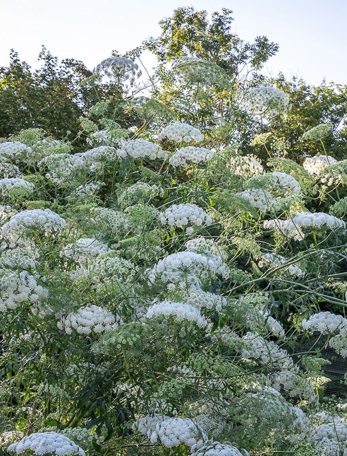
Greater Ammi
Ammi majus L.
Accepted infraspecific taxa include:
- Ammi majus subsp. majus
- Ammi majus subsp. procerum (Lowe) Menezes
Important botanical synonyms and former combinations include:
- Apium ammi Crantz, an illegitimate superfluous name
- Carum majus (L.) Koso-Pol.
- Visnaga major (L.) J.Vick
Ammi visnaga (L.) Lam., commonly called Khella or Toothpick Plant, is a separate accepted species and should not be treated as a synonym of Ammi majus.
Apiaceae Lindl. — Carrot, Parsley, or Umbel Family
Order: Apiales
Umbelliferae is the conserved historical family name commonly encountered in older botanical, medical, agricultural, and veterinary literature.
Greater Ammi; Greater Bishop’s Weed; Bishop’s Weed; Bishop’s Flower; False Bishop’s Weed; False Queen Anne’s Lace; Lady’s Lace; Laceflower; Lace Flower; White Lace Flower; White Laceflower; Bullwort; Greater Bullwort; Large Bullwort; Ammi; Aatrilal; False Khella; Falsa Biznaga; Apio Cimarrón; Berro Cimarrón; Ammi majus; Carum majus; Visnaga major; Apium ammi
“Bishop’s Weed” is highly ambiguous. It is also applied to Ground Elder, Aegopodium podagraria; the culinary spice Ajwain, Trachyspermum ammi; and sometimes the related Khella or Toothpick Plant, Ammi visnaga.
Queen Anne’s Lace ordinarily refers to Daucus carota. Greater Ammi is a separate species and should not be identified from the lace-like white flower head alone.
Ammi visnaga is a related but distinct species with denser, more rigid umbels and narrow fruiting rays historically used as toothpicks. It also contains biologically active coumarins and has its own poisoning assessment.
Regional names such as Falsa Biznaga and Cicuta Negra may create confusion with true hemlocks. Greater Ammi is not Poison Hemlock, Conium maculatum, and does not produce the same nicotinic neuromuscular-paralysis syndrome.
Confirmed Linear Furanocoumarins
The principal toxicants in Greater Ammi are phototoxic linear furanocoumarins, also called furocoumarins or psoralens. Direct chemical investigation of Texas Ammi majus seed isolated twelve linear furanocoumarins, confirming that the plant contains a chemically diverse phototoxic mixture rather than one isolated active ingredient.
Important exact-species compounds include psoralen; xanthotoxin, also called methoxsalen or 8-methoxypsoralen; bergapten, also called 5-methoxypsoralen; isopimpinellin; imperatorin; isoimperatorin; marmesin; marmesinin; and related psoralen derivatives. Fruit research has additionally identified coumarins and coumarin glucosides, including isoarnottinin and its glucosides, while umbelliprenin was isolated as a nonfuranocoumarin constituent.
Xanthotoxin and bergapten are not merely broad compounds borrowed from other Apiaceae. Both have been isolated and quantified from A. majus fruits or seeds, and the plant possesses biosynthetic machinery capable of producing methoxylated psoralens. Research cloned a Greater Ammi bergaptol O-methyltransferase involved in converting bergaptol into bergapten and showed that its activity increased strongly after biological elicitation.
Roots, Leaves, Stems, Flowers, and Reproductive Material
Fruits and their one-seeded sections are the most important concentrated exposure source. In horticultural and agricultural use, these dry fruits are commonly called seeds. Mature reproductive plants and seed-contaminated grain, hay, poultry feed, seed-cleaning waste, and supplements therefore deserve particular concern.
The hazard is not confined to mature seed. Controlled feeding of green plant material produced photosensitization in cattle and sheep, and direct skin contact with the growing plant followed by sunlight caused human photosensitization. Leaves, stems, flowers, sap, immature fruits, and crushed plant debris must therefore be treated as phototoxic.
Recent exact-species analysis also identified coumarins as the dominant positively detected chemical class in Greater Ammi roots. Xanthotoxin was isolated directly, while bergaptol-O-hexoside, isoarnottinin, and other constituents were detected through mass-spectrometric profiling. In a rat experiment, isolated xanthotoxin damaged the specialized oral mucosa and markedly reduced keratin thickness. Roots and uprooted plants should not be treated as harmless waste.
Part-specific concentration varies with genotype, maturity, plant organ, environmental conditions, stress, damage, and analytical method. Research confirms that the fruits and seeds are especially important, but no dependable concentration comparison establishes that one green leaf, flower, root, or stem is safe.
Ultraviolet Activation and DNA Injury
Linear furanocoumarins remain far more damaging when ultraviolet-A radiation activates them. These planar molecules can enter cells and position themselves between DNA base pairs. UVA exposure converts them into reactive forms capable of creating covalent bonds with pyrimidine bases, particularly thymine.
One light-dependent reaction may create a furanocoumarin–DNA monoadduct. Additional irradiation can permit a second reaction and produce an interstrand cross-link between the two DNA strands. These lesions interfere with DNA replication and transcription, impair cell division, and can initiate cell death.
Photoactivation can also generate reactive oxygen species and damage proteins, lipids, lysosomes, cell membranes, and inflammatory signaling pathways. The combined result is vascular leakage, edema, pain, blister formation, epidermal death, ulceration, and eventual tissue sloughing.
The reaction is phototoxic rather than a classic allergy. Prior sensitization is not required when sufficient plant chemical and UVA exposure occur. This explains why an animal may react severely during its first known exposure.
Systemic Primary Photosensitization
After ingestion, furanocoumarins can be absorbed and distributed to tissues exposed to light. When the animal later enters sunlight, compounds present in the skin and ocular tissues become activated. This is primary photosensitization because the plant supplies the photodynamic chemicals directly.
Primary photosensitization does not require pre-existing liver failure or impaired excretion of phylloerythrin. In a documented natural cattle outbreak, affected animals had photophobia, tearing, bilateral corneal opacity, and dermatitis of the udder, vulva, and muzzle while measured liver-associated enzyme activity remained within reference limits.
Primary disease must still be distinguished from hepatogenous photosensitization. Liver-toxic plants, mycotoxins, algae, biliary obstruction, and other hepatic disorders can permit phylloerythrin to accumulate and produce nearly identical skin lesions. Plant identification, exposure history, liver testing, and clinical pathology remain necessary.
Direct Contact and Phytophotodermatitis
Greater Ammi can also injure tissue without being swallowed. Sap, crushed foliage, flower-processing residue, fruit dust, or seed material may contaminate skin, fur, feathers, clothing, tack, tools, or bedding. Subsequent UVA exposure activates the chemicals directly within the contaminated area.
Contact lesions often preserve the exposure pattern. Streaks, drips, splashes, leaf outlines, muzzle marks, sharply edged patches, and irregular hand-shaped areas may appear where sap reached the surface. Fur can delay recognition by trapping contaminated material against the skin until later grooming or sunlight exposure.
Direct contact can coexist with systemic exposure. An animal that grazes the plant may contaminate the muzzle and face externally while simultaneously absorbing furanocoumarins through the digestive tract.
Controlled Animal Evidence and Dose Limitations
Controlled feeding studies reproduced photosensitization in cattle and sheep. In one sheep experiment, finely ground Greater Ammi seed was administered at 1, 2, 4, and 8 grams per kilogram of body weight before sunlight exposure. The lowest schedule produced mild muzzle irritation, while larger or repeated doses produced more pronounced photosensitization.
These experimental quantities document dose-related hazard but are not safe feeding thresholds. Ground seed delivered through a stomach tube differs from voluntary grazing, seed-contaminated feed, one-time ingestion, and direct sap exposure. Sun intensity, skin pigmentation, plant chemistry, dose frequency, and individual susceptibility all alter the result.
Gosling experiments demonstrated that A. majus seed was markedly photosensitizing when ingestion was followed by light exposure. Birds protected from sunlight did not develop the same characteristic reaction, confirming that the clinical syndrome depends on both the plant compounds and activating radiation.
Allergic Reactions Are a Separate Hazard
Most Greater Ammi dermatitis is nonimmune phototoxic injury. A separate occupational case documented IgE-mediated allergic rhinitis and contact urticaria in a florist handling the flowers. Immediate wheals, nasal itching, sneezing, or respiratory allergy therefore represents a different mechanism from the delayed blistering phototoxic burn.
An antihistamine may be considered by a clinician for a genuine allergic reaction, but it cannot prevent furanocoumarin photoactivation, remove the compounds from contaminated skin, or substitute for ultraviolet exclusion.
Nitrate Is Not the Defining Greater Ammi Toxin
The established species-specific poisoning syndrome is furanocoumarin photosensitization. Nitrate should not be listed beside the psoralens as though equivalent exact-species evidence proves that it routinely causes Greater Ammi poisoning.
Nitrate or nitrite poisoning may still occur in the same pasture or feed through another plant, fertilizer, or contaminated water source. Chocolate-brown blood, gray-brown or blue mucous membranes, rapid difficult breathing, and methemoglobinemia indicate a separate mechanism requiring separate testing and treatment.
Delayed Onset and Sunlight Avoidance
An exposed animal may appear normal immediately after eating or contacting Greater Ammi. Signs develop only after sufficient furanocoumarin reaches vulnerable tissue and ultraviolet radiation activates it. The delay may range from several hours to one or more days depending on dose, route, skin coverage, pigmentation, and sunlight intensity.
Early behavioral signs may be more revealing than visible skin damage. Livestock may become restless in sunlight, seek deep shade, refuse to leave a barn, shake the head, rub the face, kick at the abdomen or udder, stamp the feet, or resist handling. Horses may become difficult to lead into daylight. Dogs and cats may hide, avoid bright windows, scratch, lick, or react painfully when touched.
Cloud cover does not eliminate exposure because UVA can penetrate clouds. Bright open-sided shelters, thin tree shade, and sunlit windows may permit enough radiation to continue the reaction.
Phototoxic Dermatitis
Affected skin initially becomes warm, tender, itchy, reddened, and swollen. Edema can be pronounced around the muzzle, lips, eyelids, ears, udder, teats, vulva, scrotum, anus, and lower limbs. Pain may precede obvious discoloration.
The reaction can progress into small vesicles, large fluid-filled bullae, weeping dermatitis, crusting, fissures, erosions, ulceration, and coagulative tissue necrosis. Damaged epidermis may peel or slough in sheets, leaving raw exudative surfaces resembling a severe thermal burn.
Contact exposure may create sharply outlined streaks, drips, splashes, leaf-shaped impressions, muzzle patches, or irregular areas corresponding to contaminated fur. Systemic photosensitization more often follows the distribution of ultraviolet exposure and pigmentation.
Distribution on Unpigmented and Exposed Skin
Lesions are generally most severe on white, pink, lightly pigmented, sparsely haired, clipped, featherless, wool-free, or naturally exposed areas. Common sites include the muzzle, nose, lips, eyelids, pinnae, face, white body markings, lower legs, udder, teats, vulva, scrotum, perineum, and anus.
Melanin and dense hair or wool reduce ultraviolet penetration but do not guarantee complete protection. Severe systemic exposure, short coats, wet fur, clipped areas, scars, thin skin, and direct sap contact can permit injury outside the classic white patches.
Swelling and ulceration around the lips and muzzle may interfere with grazing, chewing, drinking, and normal rumination. Painful udder or teat lesions may prevent nursing and predispose the dam to mastitis while leaving offspring without adequate milk.
Eye and Eyelid Injury
Ocular signs include intense photophobia, excessive tearing, blinking, blepharospasm, swollen eyelids, conjunctivitis, and reluctance to open the eyes. Animals may hold the head low, seek darkness, bump into objects, or resist movement into light.
Keratitis and corneal edema can produce a blue-white or gray corneal opacity. In a documented cattle outbreak, bilateral corneal opacity, epiphora, and photophobia were prominent findings. Severe inflammation may progress to epithelial defects, ulceration, vascularization, scarring, and permanent visual impairment.
Direct seed, dust, sap, or plant-fragment exposure can add mechanical and chemical injury to systemic phototoxicity. Continuing squinting or cloudiness after irrigation requires complete ophthalmic examination rather than observation alone.
Sheep and Cattle
Sheep and cattle may develop restlessness, shade-seeking, facial edema, muzzle lesions, dermatitis of white skin, udder and vulvar injury, reduced grazing, and marked photophobia. Secondary dehydration and weight loss follow when pain prevents normal feeding and drinking.
Controlled feeding has reproduced the syndrome in both species. In the 2018 Argentine outbreak, approximately 40 percent of a 900-cow herd developed varying degrees of photophobia, tearing, bilateral corneal opacity, and dermatitis after grazing wheat stubble heavily invaded by reproductive Greater Ammi. Seeds were abundant in fecal samples, and no deaths were reported.
High morbidity with low mortality is possible when animals are recognized and removed promptly, but severe tissue loss, eye injury, secondary infection, dehydration, and delayed housing can produce a much more serious course.
Poultry and Other Birds
Goslings exposed experimentally developed photosensitization after ingesting Greater Ammi and receiving light. Reported lesions involve the poorly feathered tissues most exposed to radiation, including the beak, eyelids, face, feet, and interdigital webs.
Birds may become reluctant to stand or walk, avoid light, close the eyes, reduce feed intake, or develop swelling, crusting, ulceration, and necrosis of exposed tissues. Seed-contaminated poultry feed creates a more realistic flock hazard than casual contact with one ornamental stem.
Dense feathers offer protection over much of the body, but the beak, eyelids, comb, wattles, legs, feet, and damaged or featherless skin remain vulnerable.
Horses, Dogs, and Cats
Published exact-species cases are concentrated in livestock and poultry. Horses should nevertheless be considered susceptible to the same photodynamic mechanism and may develop painful dermatitis on white facial markings, pink noses, white lower legs, eyelids, and sparsely haired genital skin after meaningful ingestion or contact.
Dogs and cats are less likely to consume livestock-scale quantities, but garden plants, bouquets, cut stems, seed heads, and sap-contaminated fur create realistic exposures. Signs may include painful redness, swelling, licking, scratching, blistering, eye irritation, hiding, light avoidance, reduced appetite, vomiting, or diarrhea.
A shaved surgical site, thin-haired abdomen, white muzzle, pink nose, pinnae, and eyelids may be especially vulnerable. Grooming can transfer sap from the paws or coat into the mouth and eyes.
Secondary Complications
Widespread weeping or sloughed skin causes fluid, electrolyte, heat, and protein loss. Pain reduces eating, drinking, nursing, mobility, and normal social behavior. Dehydration, weakness, weight loss, and poor circulation may follow.
Open tissue permits bacterial invasion and attracts flies. Cellulitis, suppuration, fever, foul odor, spreading necrosis, fly strike, and sepsis can become more dangerous than the original photochemical reaction.
Animals repeatedly rubbing painful eyes and skin may create abrasions, self-trauma, corneal injury, or secondary wounds. Tight masks, wraps, or improvised coverings can trap heat and moisture or abrade already damaged skin.
Primary Versus Hepatogenous Photosensitization
Greater Ammi produces primary photosensitization, but outward lesions can be indistinguishable from hepatogenous disease. Animals with liver injury may accumulate phylloerythrin, a chlorophyll-derived photodynamic pigment normally excreted through bile.
Jaundice, markedly abnormal liver enzymes, bile-acid abnormalities, evidence of biliary obstruction, or exposure to hepatotoxic plants or mycotoxins supports a secondary mechanism. Normal liver-associated testing and a strong Greater Ammi exposure history support primary disease but do not eliminate every concurrent problem.
Atypical Signs and Prognosis
Chocolate-brown blood, gray-brown mucous membranes, and acute oxygen-starved collapse indicate nitrate- or nitrite-induced methemoglobinemia rather than the characteristic Greater Ammi phototoxic syndrome. Tremors, respiratory distress, seizures, or sudden death without dermatitis likewise requires investigation for another toxin or mixed exposure.
Prognosis is generally favorable when exposure is recognized before extensive tissue injury, ultraviolet light is excluded, hydration and nutrition are maintained, and the eyes remain structurally intact. Mild erythema and edema may resolve over several days.
Deep necrosis, widespread sloughing, inability to eat or nurse, severe corneal injury, secondary infection, fly strike, or delayed sunlight removal creates a guarded prognosis and may require weeks of treatment. Scarring, altered pigmentation, abnormal hair or wool growth, and lasting visual impairment may remain.
Accepted Identity, Range, and Growth Habit
Greater Ammi is Ammi majus L., an annual flowering plant in Apiaceae. Its accepted native range extends from Macaronesia and the Mediterranean region through parts of western and central Asia to southern Turkmenistan and the Arabian Peninsula.
The species has been introduced widely through ornamental cultivation, medicinal production, seed movement, contaminated agricultural material, and accidental escape. It now occurs in disturbed ground, roadsides, crop margins, waste areas, pastures, gardens, flower farms, and naturalized populations in several temperate and subtropical regions.
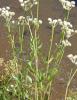
Plants may remain relatively short under dry or crowded conditions or exceed five feet where moisture, fertility, and growing conditions are favorable. One or more upright branching stems arise from a slender taproot.
Leaves, Umbels, Fruits, and Seeds
Lower leaves are broader and divided, while upper leaves become narrower and more finely dissected. The stems are generally smooth and green rather than heavily spotted or conspicuously hairy.
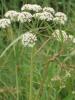
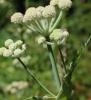
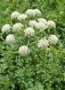
The small white flowers are arranged in broad terminal compound umbels supported by numerous rays. Divided green bracts form a conspicuous collar beneath the flower head. The entire cluster may appear flat or gently domed and creates the lace-like form valued in bouquets.
Each pollinated flower develops into a dry schizocarp that separates into two one-seeded mericarps. These fruits are commonly called seeds in horticulture, feed inspection, medicine, and toxicology. They contain some of the most important concentrations of phototoxic furanocoumarins.
Queen Anne’s Lace, Khella, and Dangerous Look-Alikes
Queen Anne’s Lace, Daucus carota, commonly has hairy stems, carrot-scented roots, deeply divided leaves, and flower heads that may contain a dark central floret and curl inward into a bird’s-nest form during fruiting. Greater Ammi is generally smoother and bears prominent divided bracts beneath a more open lace-like umbel.
Khella or Toothpick Plant, Ammi visnaga, develops denser umbels with numerous thickened rays that become rigid when the fruits mature. It is a separate species with its own coumarin chemistry and should not be substituted automatically for A. majus.
The white-flowered Apiaceae also includes Poison Hemlock, Water Hemlock, Fool’s Parsley, Wild Parsnip, Cow Parsnip, and several edible herbs. Water Hemlock can cause violent seizures and rapid death, while Poison Hemlock causes progressive neuromuscular paralysis. No white-flowered umbellifer should be identified or tasted from flower shape alone.
Experimental and Natural Animal Evidence
Feeding Greater Ammi to cattle and sheep experimentally produced photosensitization in both species. The same research documented phototoxic injury in people who handled the plant and subsequently entered sunlight, establishing that ingestion is not the only important route.
A separate sheep experiment used finely ground seed at several measured dose schedules. Mild muzzle irritation occurred at the lowest exposure, while larger and repeated exposures produced more pronounced disease after sunlight. The study demonstrates dose response but does not establish a universal safe dose for pasture plants or contaminated feed.
Comparative gosling experiments confirmed that Greater Ammi was strongly photosensitizing when seed consumption was followed by light. This evidence is especially relevant to contaminated poultry feed because birds may consume numerous fruits rapidly.
Natural cattle disease has been confirmed outside the original experimental setting. In Argentina, approximately 40 percent of a herd grazing heavily invaded wheat stubble developed ocular and cutaneous disease. Normal liver-associated results, abundant seed in feces, and the clinical distribution supported primary Greater Ammi photosensitization.
Why Light Exposure Is Essential
Plant exposure and ultraviolet exposure are separate required components of the characteristic syndrome. An animal can ingest the plant at night, remain clinically normal in enclosed housing, and become painful after entering sunlight the following day.
UVA penetrates cloud cover and can reach animals in bright open-sided shelters. Ordinary window glass blocks most UVB but can transmit a meaningful portion of UVA, so a severely exposed animal should not be placed directly beside a sunny window.
Removing an animal from the plant without removing it from ultraviolet light is therefore incomplete first aid. Likewise, moving it into shade without washing sap-contaminated fur may permit delayed injury when the animal later returns to light.
Cut Flowers, Florists, Gardeners, and Contaminated Equipment
Greater Ammi is commonly grown for wedding flowers, bouquets, and floral filler. Cut stems, discarded flower heads, seed clusters, floral buckets, processing tables, gloves, pruning tools, clothing, and delivery vehicles can all carry sap or fruit residue.
People handling the flowers have developed phototoxic dermatitis, while an occupational case also documented IgE-mediated rhinitis and contact urticaria. Waterproof gloves, long sleeves, eye protection, careful cleanup, and avoidance of sun exposure after handling are appropriate.
Pets may encounter fallen bouquet material, drink from floral buckets, walk across contaminated work surfaces, or groom sap from a person’s clothing. Floral waste should be bagged rather than left in an open trash container or compost pile accessible to animals.
Medicinal Use Does Not Establish Safety
Greater Ammi fruits have a long medicinal history because psoralen derivatives can stimulate pigmentation when combined with controlled ultraviolet exposure. Xanthotoxin contributed to the development of methoxsalen therapy and psoralen-plus-UVA treatment for selected human disorders such as vitiligo and psoriasis.
The therapeutic mechanism is the same photochemistry responsible for poisoning. Medical treatment uses characterized compounds, calculated exposure, eye protection, monitoring, and controlled irradiation. Raw fruit, homemade powder, tea, tincture, oil, poultice, or extract cannot reproduce those safeguards.
Concentrated fruit or root preparations may deliver far more furanocoumarin than casual contact with one leaf and can contain solvents, alcohol, sweeteners, or other ingredients. Greater Ammi should never be used as a homemade pigmentation, skin, parasite, reproductive, or digestive treatment for an animal.
Diagnosis and Important Differentials
There is no routine rapid veterinary assay that confirms Greater Ammi exposure in a living animal. Diagnosis depends on plant or feed identification, the timeline between exposure and ultraviolet light, lesion distribution, ocular findings, laboratory evaluation, and exclusion of secondary photosensitization.
Useful evidence includes complete-plant photographs, flower and fruit heads, representative leaves and stems, nursery or seed labels, bouquet material, hay, grain, poultry feed, fecal fruits, and information about when the animal entered sunlight.
A complete blood count, proteins, electrolytes, kidney values, liver enzymes, bilirubin, bile acids, and urinalysis may be appropriate according to severity. Liver evaluation helps distinguish primary furanocoumarin disease from hepatogenous phylloerythrin photosensitization.
Eye examination may require fluorescein staining, tear evaluation, eyelid eversion, magnified corneal inspection, and intraocular-pressure measurement. Skin culture, cytology, or biopsy may be indicated for infected, necrotic, atypical, or poorly healing lesions.
Nitrate testing is appropriate only when forage, water, fertilizer, weather, or clinical findings independently support that concern. Chocolate-brown blood and methemoglobinemia do not explain sunlight-dependent blistering and corneal opacity.
Prevention and Prognosis
Remove Greater Ammi from animal areas before the reproductive stage concentrates fruit and seed. Cut plants should be collected immediately; wilting or drying does not instantly eliminate furanocoumarins.
Inspect hay, grain, poultry feed, seed-cleaning waste, bedding, and supplements for fruit contamination. Do not release hungry livestock into a dense stand, and do not place flower-farm or garden waste in paddocks, poultry runs, rabbit enclosures, kennels, or open compost.
Most animals recover when the source is removed, ultraviolet exposure is stopped early, and skin, eye, hydration, nutrition, infection, and fly control are managed effectively. Extensive necrosis or corneal disease may require prolonged treatment and can leave permanent cosmetic or visual consequences.
Immediate Ultraviolet Protection
- Move the animal out of ultraviolet light immediately. Use dim enclosed housing or complete opaque shelter rather than thin tree shade or an open-sided structure.
- Do not wait for lesions. Redness, swelling, blistering, and corneal opacity may develop hours after the plant has been eaten or removed.
- Protect during transport. Close or cover sun-facing trailer openings when ventilation can be maintained, or transport after dark when practical.
- Limit bright-window exposure. Keep severely exposed companion animals away from direct sunlight passing through windows.
- Continue restriction until veterinary release. Returning outside too early can initiate new injury or extend lesions while photodynamic compounds remain in tissue.
Remove Plant Residue Safely
- Wear protective equipment. Use waterproof gloves, long sleeves, long pants, and eye protection before touching the plant, contaminated fur, bedding, tack, or equipment.
- Prevent grooming and rubbing. Stop dogs and cats from licking contaminated fur and prevent livestock from rubbing painful skin or eyes.
- Lift away visible material. Remove leaves, stems, flowers, fruits, seeds, or plant fragments without crushing them against the skin.
- Wash skin and fur thoroughly. Use mild soap or pet-safe shampoo with cool or lukewarm water. Rinse completely and direct runoff away from the face and previously clean skin.
- Repeat gentle washing when necessary. Heavy sap or seed-dust contamination may require a second wash with fresh gloves and clean water.
- Do not scrub aggressively. Vigorous friction can damage the surface and spread sap across a larger area.
- Clean contaminated objects. Wash collars, harnesses, halters, blankets, brushes, buckets, tools, boots, clothing, vehicles, stalls, and flower-processing surfaces.
Eye Exposure
Begin irrigation immediately with sterile saline or clean lukewarm water. Flush continuously for at least 15 to 20 minutes, allowing fluid to carry plant residue away from the eye and unaffected side of the face.
- Do not rub or wipe the cornea. Seeds, dust, and fragments can abrade the surface.
- Prevent pawing and face rubbing. Use safe restraint or an Elizabethan collar when available.
- Maintain dim lighting. Photophobia can be intense even before cloudiness becomes visible.
- Seek prompt examination. Direct eye contamination, persistent squinting, tearing, eyelid swelling, redness, cloudiness, or visual uncertainty requires veterinary evaluation.
- Do not use leftover eye medication. Human redness-relief drops and old veterinary products may be inappropriate. Steroid-containing eye medication can worsen an undiagnosed corneal ulcer.
Veterinary examination may include fluorescein staining, tear testing, eyelid eversion, corneal magnification, pressure measurement, lubrication, pain control, and topical antimicrobial treatment when an epithelial defect or ulcer is present. Ophthalmic corticosteroids are inappropriate until corneal ulceration has been excluded.
Do Not Attempt Unsupervised Home Decontamination
Do not automatically induce vomiting. The appropriate decision depends on species, plant part, amount, timing, neurologic condition, airway protection, and whether the exposure involved concentrated fruits or seeds.
- Do not use hydrogen peroxide, salt, mustard, ipecac, detergent, dish soap, or manual gagging.
- Never use hydrogen peroxide as a feline emetic.
- Do not induce vomiting in livestock, horses, rabbits, guinea pigs, birds, or other species incapable of safe emesis.
- Do not induce vomiting in a symptomatic animal. Weakness, repeated vomiting, respiratory abnormalities, poor coordination, seizures, collapse, or impaired swallowing creates aspiration risk.
- Do not force activated charcoal. A veterinarian may consider it after a substantial recent fruit or seed ingestion only when the animal can protect its airway.
- Do not administer routine cathartics. They can worsen dehydration and electrolyte loss.
- Do not give milk, oil, bread, herbs, household charcoal, vitamins, antihistamines, corticosteroids, or other supposed antidotes.
When Emergency Examination Is Especially Important
- Eye pain or opacity: Severe squinting, inability to open the eye, blue-white cloudiness, apparent blindness, or progressive eyelid swelling requires urgent ophthalmic care.
- Rapidly expanding dermatitis: Increasing edema, blistering, weeping, ulceration, or skin separation indicates active tissue injury.
- Extensive white-skin involvement: Large affected areas can cause major pain, fluid loss, and secondary infection.
- Inability to eat, drink, graze, or nurse: Facial, oral, udder, or teat lesions can rapidly produce dehydration and nutritional compromise.
- Fever, foul odor, pus, or spreading redness: These signs suggest secondary bacterial infection.
- Fly strike: Eggs or larvae in wounds require immediate treatment.
- Weakness, collapse, gray-brown mucous membranes, or abnormal breathing: Another toxin, nitrate exposure, severe dehydration, or systemic complication may be present.
Veterinary Assessment
The veterinarian will assess the route and timing of exposure, duration of ultraviolet exposure, total skin area involved, lesion depth, hydration, temperature, pain, feeding ability, nursing, ocular injury, secondary infection, and evidence of another photosensitization mechanism.
Blood testing may include a complete blood count, total protein, albumin, electrolytes, glucose, kidney values, liver enzymes, bilirubin, and bile acids. Normal liver-associated testing supports primary photosensitization, whereas significant hepatic or biliary abnormalities may indicate a secondary mechanism.
Skin cytology, bacterial culture, or biopsy may be appropriate for infected, necrotic, unusual, or nonhealing lesions. Feed, fruit, seed, hay, and pasture samples should be preserved for botanical or chemical analysis.
Pain, Inflammation, and Wound Care
Phototoxic dermatitis can be intensely painful. Veterinarian-selected systemic analgesia is often necessary, particularly when lesions involve the muzzle, eyelids, feet, udder, teats, or large body areas.
Anti-inflammatory treatment may be considered early in selected cases, but no medication reverses DNA photoadducts or makes continued sunlight safe. Corticosteroids and nonsteroidal anti-inflammatory drugs require patient-specific assessment because dehydration, infection, pregnancy, gastrointestinal disease, and species differences alter their safety.
Intact blisters should not be deliberately opened at home. Open wounds may be cleaned with sterile saline or another veterinarian-selected solution and protected with nonadherent dressings when appropriate. Some lesions heal better uncovered in a clean indoor environment; bandaging decisions depend on location, moisture, depth, and the animal.
Do not apply peroxide, alcohol, bleach, concentrated iodine, essential oils, solvents, human burn cream, topical anesthetics, zinc-containing ointments, or fragranced products without approval. Products harmless to human skin may be toxic when licked or may trap heat and contamination.
Fluids, Nutrition, and Extensive Skin Loss
Animals with large exudative lesions, reduced intake, vomiting, diarrhea, heat exposure, or fever may require intravenous fluids and electrolyte correction. Protein loss through widespread damaged skin can contribute to edema, weakness, and delayed healing.
Soft or modified feed may be needed when muzzle or lip lesions make chewing painful. Assisted feeding should be performed only when swallowing is safe. Nursing offspring may require temporary supplemental feeding when udder or teat pain prevents normal access.
Temperature, body weight, hydration, urine output, blood proteins, appetite, and lesion progression may require repeated monitoring during severe cases.
Infection and Fly-Strike Control
House affected animals in a clean, dry, well-ventilated, dim, insect-controlled environment. Replace wet bedding and remove exudate-contaminated materials promptly.
Antibiotics are appropriate when bacterial infection is present or strongly suspected rather than automatically for every phototoxic lesion. Culture can guide treatment in deep, foul-smelling, spreading, or poorly responsive infections.
Inspect beneath crusts, wool, feathers, skin folds, sheets, masks, wraps, and bandages for heat, odor, discharge, eggs, or larvae. Fly strike can progress rapidly in warm weather and may require clipping, lavage, larval removal, debridement, systemic treatment, and aggressive supportive care.
Livestock insecticides and repellents should not be placed directly on open tissue unless the veterinarian approves the exact product and species use.
Livestock and Horse Management
Remove the entire exposed herd or flock from Greater Ammi, contaminated stubble, hay, grain, seed waste, or clippings. Individual consumption and lesion onset may differ, so clinically normal animals require continued observation.
Inspect white faces, pink noses, eyelids, ears, udders, teats, vulvas, scrotums, perineal skin, white lower legs, and sparsely haired areas at least several times daily. Examine the eyes for tearing, squinting, opacity, and impaired navigation.
Provide uncontaminated forage and clean water inside the protected housing. Do not force affected animals to exercise. Night grazing may be considered only after the source has been removed and the veterinarian determines that outdoor access, fencing, wound protection, and monitoring are adequate.
Dogs and Cats
Keep exposed pets in a dim interior room away from sunny windows. Prevent grooming with an Elizabethan collar when sap may remain on the coat, and examine the muzzle, nose, lips, eyelids, pinnae, abdomen, groin, inner limbs, and any clipped areas.
Monitor appetite, drinking, vomiting, diarrhea, skin pain, swelling, blistering, squinting, and light avoidance. Delayed lesions require repeated checks during the following several days even when the pet initially appears normal.
Nitrate Remains a Separate Differential
Methylene blue is not an antidote for Greater Ammi furanocoumarins. It does not prevent photoactivation, repair DNA cross-links, treat corneal damage, or stop phototoxic dermatitis.
A veterinarian may use methylene blue for confirmed or strongly suspected nitrate- or nitrite-induced methemoglobinemia. Chocolate-brown blood, gray-brown or blue mucous membranes, severe oxygen-starved breathing, and high measured methemoglobin warrant investigation of forage, water, fertilizer, and other plants rather than automatic attribution to Greater Ammi.
Recovery and Prognosis
Mild erythema, edema, and discomfort may improve over several days with prompt washing and strict ultraviolet protection. Blisters, ulcers, and superficial tissue loss may require several weeks of care.
Deep necrosis can slough gradually and may leave permanent scarring, altered pigmentation, or abnormal hair, feather, or wool growth. Corneal ulcers and stromal injury can require prolonged treatment and may leave lasting opacity or visual impairment.
Ultraviolet restriction must continue until the veterinarian determines that the animal can return gradually without renewed pain or lesion progression. A single premature outdoor exposure may reactivate injury.
Frequently Asked Questions About Greater Ammi and Animal Photosensitization
Can an animal appear normal for hours after eating Greater Ammi?
Yes. Furanocoumarins require ultraviolet activation. An animal may ingest the plant, remain comfortable in enclosed housing, and develop pain, swelling, photophobia, or blistering only after entering sunlight. This delay is why ultraviolet exclusion should begin immediately rather than after visible lesions appear.
Does normal liver testing rule out photosensitization?
No. Normal liver-associated results are compatible with primary Greater Ammi photosensitization because the plant supplies the photodynamic chemicals directly. Liver testing is useful because it helps distinguish primary disease from hepatogenous photosensitization caused by impaired phylloerythrin excretion.
Why can the cornea turn blue-white or cloudy?
Phototoxic inflammation can damage the corneal epithelium and disrupt normal fluid regulation within the cornea, producing edema and opacity. Direct seed, dust, or sap exposure may add abrasion or chemical injury. Persistent cloudiness requires examination because ulceration and deeper stromal damage can threaten vision.
Are dried fruits, seeds, hay, and cut flowers still hazardous?
Yes. Drying does not immediately destroy the linear furanocoumarins. Mature fruits can contaminate feed, grain, hay, floral waste, and seed-cleaning residue, while cut stems and dried flower material can retain phototoxic compounds. Dried material also produces dust that can contaminate skin and eyes.
Can contaminated fur cause injury after the plant has been removed?
Yes. Sap or fruit residue trapped in fur can remain against the skin and may be transferred to the mouth or eyes during grooming. Injury may occur later when the animal enters ultraviolet light. Washing the coat and cleaning contaminated collars, bedding, tack, and equipment are therefore part of treatment.
Is a dark-coated or dark-skinned animal completely protected?
No. Melanin and dense hair reduce ultraviolet penetration, so white or sparsely covered areas are usually injured most severely. Direct sap contact, clipped skin, scars, thin hair, eyelids, lips, and extensive systemic exposure can still affect a pigmented animal.
Can cloudy weather or ordinary shade activate the toxins?
Yes. Ultraviolet-A radiation penetrates cloud cover and can reach an animal beneath thin trees or inside an open-sided shelter. Strong sunlight through windows may also provide UVA exposure. Dim enclosed housing or genuinely opaque shelter is safer during the active period.
Is allergic contact urticaria the same as a Greater Ammi phototoxic burn?
No. Phototoxic dermatitis is a nonimmune reaction requiring furanocoumarin contact or absorption followed by ultraviolet exposure. Contact urticaria is an immediate allergic reaction that may produce wheals and itching without the same delay. A florist has developed IgE-mediated rhinitis and contact urticaria from Greater Ammi, so both mechanisms are possible.
Why are reproductive plants and contaminated feed especially dangerous?
The fruits and seeds contain concentrated mixtures of linear furanocoumarins and can be eaten rapidly in grain, hay, poultry feed, crop residue, or seed-cleaning waste. In a natural cattle outbreak, numerous Greater Ammi seeds were found in feces from affected animals grazing reproductive plants.
Are Greater Ammi and Khella the same plant?
No. Greater Ammi is Ammi majus. Khella or Toothpick Plant is Ammi visnaga. Both contain biologically active coumarins and can photosensitize animals, but they are separate species with different morphology, compound profiles, and research records.
Can sunscreen make it safe to return an affected animal outdoors?
No. Sunscreen cannot guarantee complete coverage, may be removed by licking or rubbing, and may contain ingredients unsafe for the species or open wounds. Physical ultraviolet exclusion remains the primary protection until a veterinarian approves gradual return outdoors.